. 2006 Jun;168(6):1879-88.
doi: 10.2353/ajpath.2006.051170.

Isolation of a novel population of multipotent adult stem cells from human hair follicles

Affiliations

Isolation of a novel population of multipotent adult stem cells from human hair follicles

Hong Yu et al. Am J Pathol. 2006 Jun.

Abstract

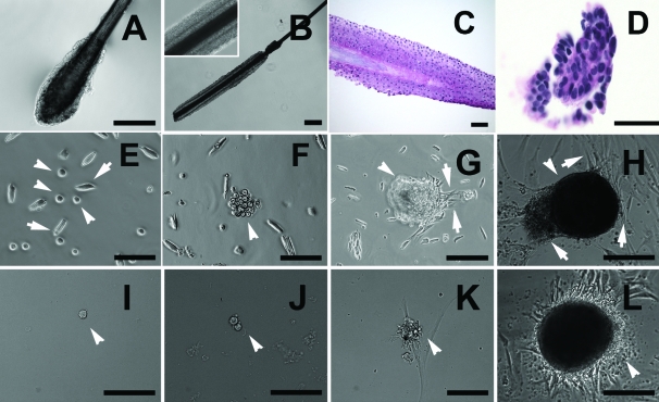
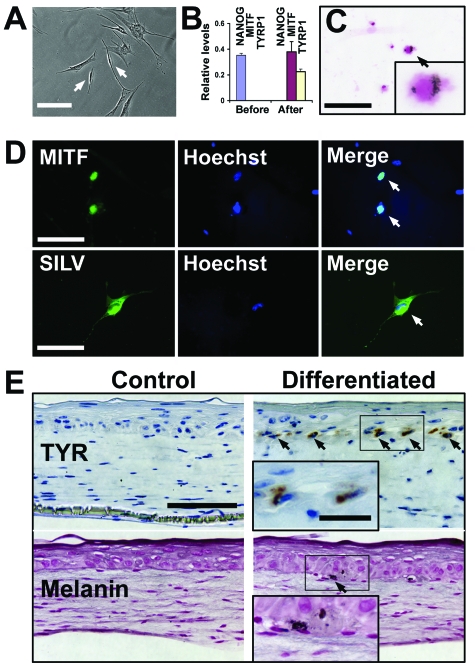
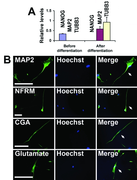
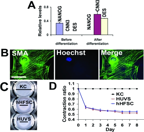
Hair follicles are known to contain a well-characterized niche for adult stem cells: the bulge, which contains epithelial and melanocytic stem cells. Using human embryonic stem cell culture conditions, we isolated a population of adult stem cells from human hair follicles that are distinctively different from known epithelial or melanocytic stem cells. These cells do not express squamous or melanocytic markers but express neural crest and neuron stem cell markers as well as the embryonic stem cell transcription factors Nanog and Oct4. These precursor cells proliferate as spheres, are capable of self-renewal, and can differentiate into multiple lineages. Differentiated cells not only acquire lineage-specific markers but also demonstrate appropriate functions in ex vivo conditions. Most of the Oct4-positive cells in human skin were located in the area highlighted by cytokeratin 15 staining in vivo. Our data suggest that human embryonic stem cell medium can be used to isolate and expand human adult stem cells. Using this method, we isolated a novel population of multipotent adult stem cells from human hair follicles, and these cells appear to be located in the bulge area. Human hair follicles may provide an accessible, autologous source of adult stem cells for therapeutic application.


Figures

Figure 1
Figure 1
Isolation and expansion of hair spheres in hESC medium. A: Morphology of a plucked catagen/telogen hair follicle. B: Morphology of a plucked anagen hair follicle. C: Histology of a plucked anagen hair follicle (H&E stain). D: Histology of a plucked catagen/telogen hair follicle (H&E). E: A suspension of single cells, obtained from trypsinized follicular epithelium, contains a heterogeneous cell population. Some cells display relatively large spindle morphology (arrows), whereas others are small round cells (arrowheads). F: Small round cells proliferate to form a cell cluster in suspension in hESC medium (arrowheads). G and H: Cell clusters become adherent to the culture plates and form sphere-like structures (arrowheads) with sprouting, adherent fibroblastic cells at the periphery (arrows). IL: A single cell derived from a hair sphere recapitulates the similar process and re-forms a sphere with adherent fibroblastic cells at the periphery (arrowheads). Representative images were taken from at least three independent experiments. Scale bars in A, B, C, D, and G = 200 μm; in E, F, H, I, J, K, and L = 100 μm.
Figure 2
Figure 2
Human hair follicle-derived stem cells express various stem cell markers. A: Immunocytochemistry and confocal microscopy reveals expression of Nanog (red) and Oct4 (green) in a portion of heterogeneous populations within hair spheres. Their expression was typically localized to nuclei (stained blue with To Pro 3) (arrowheads). Nestin shows a cytoplasmic staining pattern. B: Spheres were dissociated into single cells and plated onto Matrigel-coated chamber slides for staining. Confocal images show that individual cells display nuclear staining pattern for Nanog (red) and Oct4 (green); cytoplasmic staining for Nestin. A characteristic punctate pattern of Oct4 staining is observed in both small round cells (top) and intermediate to large cells (bottom, arrows). The latter show a relatively weak staining intensity. Nanog immunoreactivity is observed in small round cells (bottom) and a portion of intermediate sized cells (top, arrows). Nestin also stains spindled cells (bottom). C: Real-time RT-PCR analysis shows that hHFSCs expressed SNAIL, SLUG, SOX9, TWIST, nestin (NES), and BMP4 genes. We used epidermal keratinocytes (KC) and embryonic bodies derived from hESCs as negative and positive controls, respectively. Representative images were taken from at least three independent experiments. Data shown are mean ± SD from three independent experiments. Scale bars in A for Oct4 = 18.75 μm and for Nanog and Nestin = 75 μm; and in B for Oct4: top = 3.75 μm, bottom = 30 μm; for Nanog: top = 7.5 μm, bottom = 30 μm; and for Nestin: top = 7.5 μm, bottom = 30 μm.
Figure 3
Figure 3
Melanocytic differentiation of hHFSCs. A: Dissociated, hair sphere-derived cells were treated in melanocyte differentiation medium for 14 days. We observed dendritic cells with typical melanocyte morphology (arrows). B: MITF and TYRP1 gene expression is detected after differentiation; however, expression of stemness gene NANOG is undetectable after differentiation. C: Fontana-Mason staining shows melanin pigment in a portion of differentiated cells (arrow and inset). D: Differentiated cells exhibit nuclear and cytoplasmic immunoreactivity for MITF and SILV/HMB45, respectively. E: After embedding in three-dimensional human skin reconstructs, differentiated cells homed to the dermal-epidermal junction (arrows and insets). They are readily identifiable by TYR immunoreactivity or by Fontana-Mason staining. Reconstructs embedded with undifferentiated hFSCs do not show TYR and melanin staining. Representative images were taken from two independent experiments. Scale bars in A and D (SILV) = 200 μm; in B, D (MITF), and E = 100 μm; and in inset in E = 30 μm.
Figure 4
Figure 4
Neural differentiation of hHFSCs. A: Real-time RT-PCR demonstrates expression of neuronal markers MAP2 and β3-tubulin (TUBB3) and loss of NANOG after neuronal differentiation. B: Differentiated cells become immunoreactive for MAP2, NFM, CGA, and neurotransmitter glutamate. MAP2 preferentially stains dendritic processes versus cell bodies (arrow). NFM, CGA, and glutamate stain cytoplasm of neuron-differentiated cells (arrows). Representative images were taken from at least three independent experiments. Scale bars in A = 100 μm.
Figure 5
Figure 5
Smooth muscle cell differentiation of hHFSCs. A: Real-time RT-PCR demonstrates expression of calponin (CNN3) and desmin (DES) and loss of NANOG after smooth muscle differentiation. B: Differentiated cells become immunoreactive for SMA. SMA stains cytoplasm of differentiated smooth muscle cells. C: Contraction assays show hHFSC contracted collagen gels. Epidermal keratinocytes (KC) and HUVSs serve as negative and positive controls, respectively. Black bars indicate collagen gel diameters. D: Quantification of collagen gel shrinkage shows that differentiated cells contract collagen gel similarly to HUVS control. Data shown are mean ± SD of contraction ratio relative to keratinocyte control from three independent experiments. Scale bars in A = 100 μm.
Figure 6
Figure 6
Oct4-positive cells are resided in the hair bulge. A: Oct4 and K15 immunohistochemical stains on consecutive normal human scalp tissues. Top: An anagen hair follicle; bottom: a catagen/telogen hair follicle. K15 highlights the bulge area, and a few Oct4-positive cells are present within K15-highlighted areas. B: Individual hair follicles were cultured in hESC medium for 6 days and then stained with Oct4 and Nanog. Immunostaining and differential interference contrast (DIC) images show that catagen/telogen bulge contains OCT4-positive cells (green, arrows) or NANOG-positive cells (red, arrows). C: Confocal images of catagen/telogen bulges reveal nuclear localization of OCT4 (green, arrow) and Nanog (red, arrow) in the bulge cells. Nuclei are counterstained with TO-PRO-3 (blue). Representative images were taken from at least three independent experiments. Scale bars in A = 30 μm; in B = 200 μm; and in C = 18.75 μm.

References

    1. Bjornson CR, Rietze RL, Reynolds BA, Magli MC, Vescovi AL. Turning brain into blood: a hematopoietic fate adopted by adult neural stem cells in vivo. Science. 1999;283:534–537. - PubMed
    1. Clarke DL, Johansson CB, Wilbertz J, Veress B, Nilsson E, Karlstrom H, Lendahl U, Frisen J. Generalized potential of adult neural stem cells. Science. 2000;288:1660–1663. - PubMed
    1. Mezey E, Chandross KJ, Harta G, Maki RA, McKercher SR. Turning blood into brain: cells bearing neuronal antigens generated in vivo from bone marrow. Science. 2000;290:1779–1782. - PubMed
    1. Jiang Y, Jahagirdar BN, Reinhardt RL, Schwartz RE, Keene CD, Ortiz-Gonzalez XR, Reyes M, Lenvik T, Lund T, Blackstad M, Du J, Aldrich S, Lisberg A, Low WC, Largaespada DA, Verfaillie CM. Pluripotency of mesenchymal stem cells derived from adult marrow. Nature. 2002;418:41–49. - PubMed
    1. Orlic D. Adult bone marrow stem cells regenerate myocardium in ischemic heart disease. Ann NY Acad Sci. 2003;996:152–157. - PubMed

Publication types

MeSH terms