. 2006 Jun;168(6):1910-20.
doi: 10.2353/ajpath.2006.050636.

Toxoplasma gondii infection reveals a novel regulatory role for galectin-3 in the interface of innate and adaptive immunity

Affiliations

Toxoplasma gondii infection reveals a novel regulatory role for galectin-3 in the interface of innate and adaptive immunity

Emerson Soares Bernardes et al. Am J Pathol. 2006 Jun.

Abstract

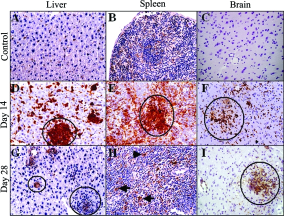
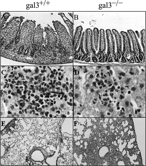
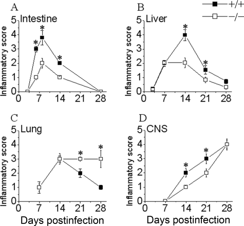
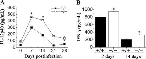
In attempts to investigate the role of galectin-3 in innate immunity, we studied galectin-3-deficient (gal3-/-) mice with regard to their response to Toxoplasma gondii infection, which is characterized by inflammation in affected organs, Th-1-polarized immune response, and accumulation of cysts in the central nervous system. In wild-type (gal3+/+) mice, infected orally, galectin-3 was highly expressed in the leukocytes infiltrating the intestines, liver, lungs, and brain. Compared with gal3+/+, infected gal3-/- mice developed reduced inflammatory response in all of these organs but the lungs. Brain of gal3-/- mice displayed a significantly reduced number of infiltrating monocytes/macrophages and CD8+ cells and a higher parasite burden. Furthermore, gal3-/- mice mounted a higher Th1-polarized response and had comparable survival rates on peroral T. gondii infection, even though they were more susceptible to intraperitoneal infection. Interestingly, splenic cells and purified CD11c+ dendritic cells from gal3-/- mice produced higher amounts of interleukin-12 than cells from gal3+/+ mice, possibly explaining the higher Th1 response verified in the gal3-/- mice. We conclude that galectin-3 exerts an important role in innate immunity, including not only a pro-inflammatory effect but also a regulatory role on dendritic cells, capable of interfering in the adaptive immune response.


Figures

Figure 1
Figure 1
Immunohistochemical staining for galectin-3 in organs of T. gondii-infected wild-type mice. The photomicrographs depict galectin-3 immunostaining (brown color) with a hematoxylin counterstain. In the uninfected mice, few cells in the liver (A) and spleen (B) express galectin-3, and galectin-3 staining is barely noted in the brain (C). On day 14 after T. gondii infection, galectin-3 is expressed by a high number of inflammatory cells in the liver (D), spleen (E), and brain (F). On day 28 after T. gondii infection, the level of galectin-3 expression in the liver (G) and spleen (H) decline to almost basal levels but remain high in the brain (I). All photomicrographs are under magnification with ×40 objective. Circles show inflammatory foci, and arrows point to galectin-3-expressing cells.
Figure 2
Figure 2
Histopathology and inflammatory response in tissues of T. gondii-infected gal3+/+ and gal3−/− mice. Mice were infected with 20 cysts of the ME-49 strain of T. gondii, and histological studies were performed on their intestine, liver, and lung at several intervals after infection. A and B: The intestines of gal3+/+ and gal3−/− mice, respectively, show initial stages of tissue necrosis accompanied by an increased number of inflammatory cells in the lamina propria and submucosa on day 6 (A). In contrast, gal3−/− mice exhibited a preserved villous architecture on day 6 (B). C and D: The liver of gal3+/+ and gal3−/− mice, respectively, on day 14 after infection. E and F: The lung of gal3+/+ and gal3−/− mice, respectively, on day 28 after infection. Stronger inflammatory infiltration persists in the lung of gal3−/−. H&E; A, B, E, and F, under magnification with ×10 objective; C and D, under magnification with ×60 objective.
Figure 3
Figure 3
Summary of the inflammatory response kinetics in the peripheral organs and CNS of T. gondii-infected gal3+/+ and gal3−/− mice. A (intestine), B (liver), C (lung), and D (CNS) show the evolution of inflammation intensity at several intervals after infection. The inflammatory scores were measured as described in Materials and Methods. Asterisks indicate that differences are statistically significant at P < 0.05.
Figure 4
Figure 4
Parasite burden in the brain of T. gondii-infected gal3+/+ and gal3−/− mice. The mice were perorally infected with 20 cysts of the ME-49 strain of T. gondii. A: Arrows and ellipse indicate cysts in the brains of gal3+/+ and gal3−/− mice on day 14 after infection (parasite-specific immunostaining; magnification with ×40 objective). B: The number of cysts detected by immunocytochemistry in brain sections of T. gondii-infected gal3+/+ and gal3−/− mice during the course of the infection. *Significantly different from values obtained with the gal3+/+ mice (P < 0.03). Similar results were obtained in two experiments. C and D: The numbers of infiltrating CD4+, CD8+, and F4/80+ cells into the brains of T. gondii-infected gal3+/+ and gal3−/− mice 14 and 28 days after infection, respectively. Cells were counted by immunohistochemistry, and data represent the mean ± SEM, *P < 0.05.
Figure 5
Figure 5
Survival of gal3+/+ and gal3−/− mice to T. gondii infection. Mice were perorally (A) or intraperitoneally (B) infected with 20 cysts of the ME-49 strain of T. gondii, and survival was monitored. Data are representative of three experiments, each performed with five to eight mice per group, yielding similar results. The results are expressed as percentage of live animals during the course of infection. A: No difference in mortality was observed between gal3+/+ and gal3−/− after oral infection. B: The gal3−/− mice infected by the intraperitoneal route exhibit significantly lower survival than gal3+/+ mice infected by the same route (P < 0.027).
Figure 6
Figure 6
Reduced recruitment of inflammatory monocytes/macrophages and neutrophils, and high parasite burden in gal3−/− mice after intraperitoneal infection with T. gondii. Peritoneal cells from gal3+/+ and gal3−/− mice were harvested 4 days after intraperitoneal inoculation of a dose of 20 cysts of the ME-49 strain of T. gondii, and subpopulations of leukocytes in the fluid were enumerated. A shows the recoveries for neutrophils and B for macrophages 4 days after infection (pi). C: Shows cyst numbers counted from whole brain homogenates 14 days after infection (pi). Results shown are representative of individual mice. The experiment was repeated twice with similar results, using a total of five mice per genotype. *P < 0.03.
Figure 7
Figure 7
Levels of IL-12p40 and IFN-γ and ratio of parasite-specific IgG2a to IgG1-isotype in the sera of T. gondii-infected mice. The serum samples were collected on day 14 after infection. IL-12p40 (A), IFN-γ (B), and IgG2a/IgG1 (C) levels were assayed by ELISA. The results represent the mean ± SD of data from five mice of each genotype. *P < 0.03. A: The difference between the responses obtained with the gal3+/+ and gal3−/− mice are significantly different throughout the entire period (P < 0.05). Similar results were obtained in three experiments.
Figure 8
Figure 8
Levels of IL-12p40 in the supernatants of spleen cell from T. gondii-infected mice. Spleen cells, harvested on days 7, 14, 21, and 28 after infection, were incubated with medium alone for 48 hours. The supernatants were collected and assayed for IL-12p40 (A) and IFN-γ by ELISA. A: The results represent the mean ± SD of triplicate cultures from a representative experiment. *P < 0.02. The difference between the responses obtained with the gal3+/+ and gal3−/− mice are significantly different throughout the entire period (P < 0.05). B: Levels of IFN-γ in the supernatant of the spleen cells harvested on days 7 and 14 after infection, without any re-stimulation. After day 15, IFN-γ in the supernatant is below the detection limit in all groups, unless STAg stimulation is used. Results are reported as the mean ± SD. *P < 0.05. The experiment was repeated a second time with similar results.
Figure 9
Figure 9
Production of IL-12p40, IL12p70, and IFN-γ by the spleen cells from uninfected gal3+/+ and gal3−/− mice. Total cells, macrophages, and CD11c+ dendritic cells isolated from the spleen of uninfected gal3+/+ and gal3−/− mice were cultured for 48 hours in the presence or absence of LPS, IFN-γ, or LPS + IFN-γ. The concentrations of IFN-γ (A), IL-12p40 (B), and IL-12p70 (C) in the culture supernatants were measured by ELISA. The spleen cells of gal3−/− mice produced higher levels of IFN-γ (A), IL-12p40 (B), and IL-12p70 (C) when compared with those of gal+/+ mice. CD11c+ dendritic cells but not macrophages account for the higher levels of IL-12p40 in gal3−/− mice (D) after stimulation with LPS + IFN-γ. The results represent the mean ± SD of triplicate cultures from a representative experiment. *P < 0.02.

References

    1. Barondes SH, Cooper DNW, Gitt MA, Leffler H. Galectins: structure and function of a large family of animal lectins. J Biol Chem. 1994;269:20807–20810. - PubMed
    1. Kasai K, Hirabayashi J. Galectins: a family of animal lectins that decipher glycocodes. J Biochem. 1996;119:1–8. - PubMed
    1. Hughes RC. The galectin family of mammalian carbohydrate-binding molecules. Biochem Soc Trans. 1997;25:1194–1198. - PubMed
    1. Leffler H, Carlsson S, Hedlund M, Qian Y, Poirier F. Introduction to galectins. Glycoconj J. 2004;19:433–440. - PubMed
    1. Liu FT. Regulatory roles of galectins in the immune response. Int Arch Allergy Immunol. 2005;136:385–400. - PubMed

Publication types