. 2006 Jun;168(6):1960-74.
doi: 10.2353/ajpath.2006.051113.

Multiorgan autoimmune inflammation, enhanced lymphoproliferation, and impaired homeostasis of reactive oxygen species in mice lacking the antioxidant-activated transcription factor Nrf2

Affiliations

Multiorgan autoimmune inflammation, enhanced lymphoproliferation, and impaired homeostasis of reactive oxygen species in mice lacking the antioxidant-activated transcription factor Nrf2

Qiang Ma et al. Am J Pathol. 2006 Jun.

Abstract

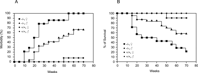
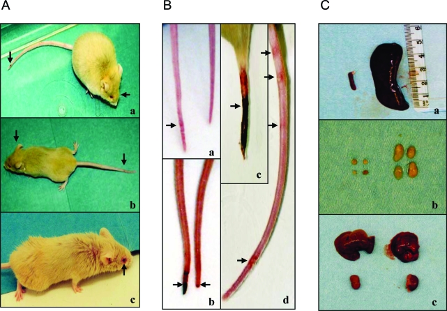
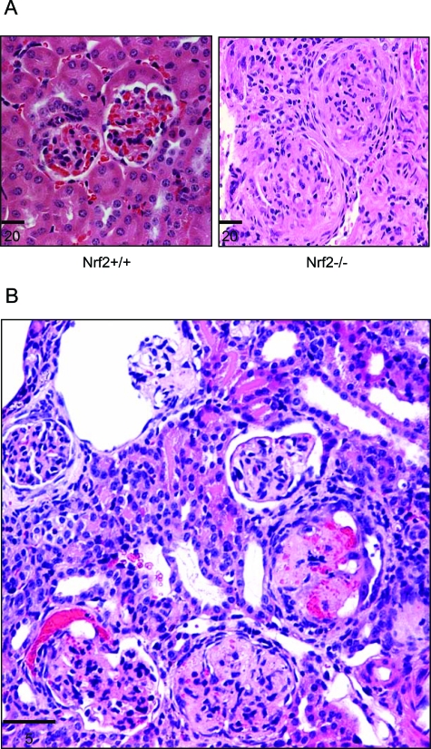
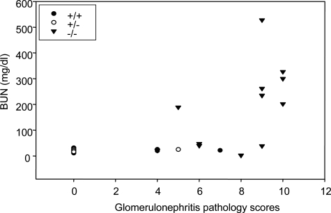
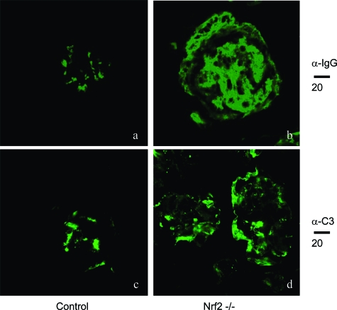
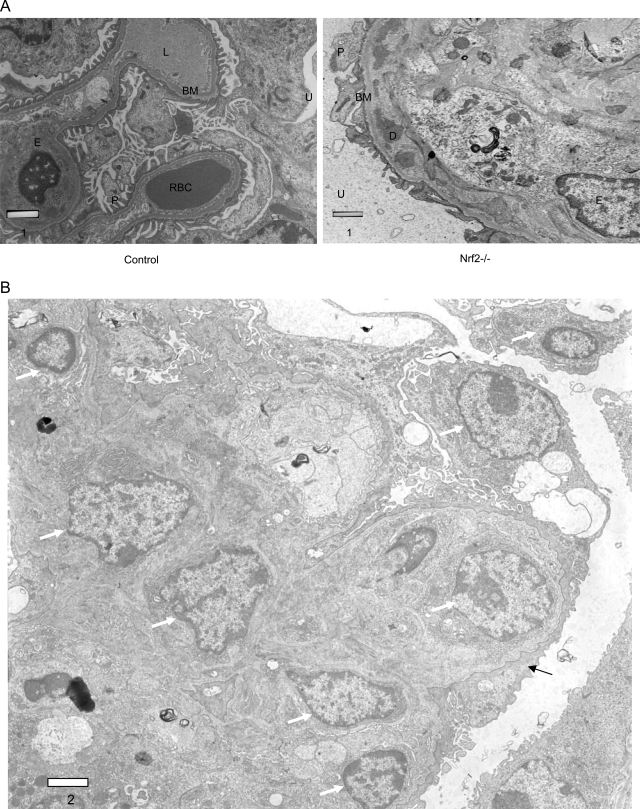
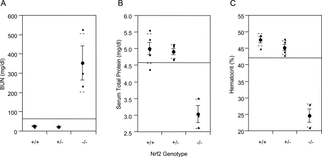
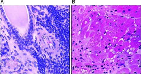
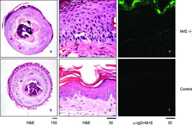
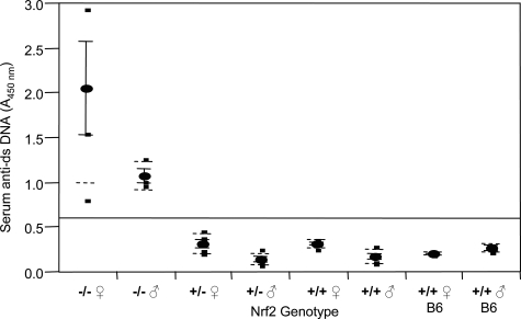
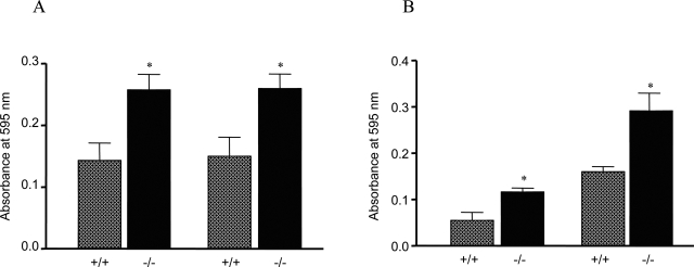
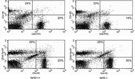
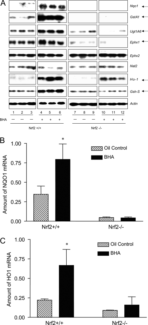
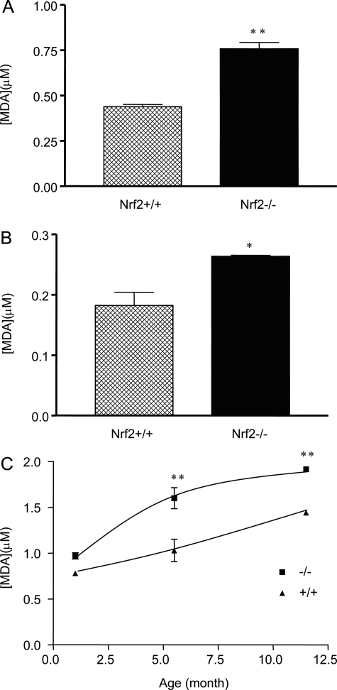
Nuclear factor erythroid 2-related factor 2 (Nrf2) is an antioxidant-activated cap "n" collar basic leucine zipper transcription factor. To assess the function of Nrf2 in the antioxidant response, we examined mice with targeted disruption of the Nrf2 gene. Nrf2-null mice developed complex disease manifestations, with a majority exhibiting a lupus-like autoimmune syndrome characterized by multiorgan inflammatory lesions with a marked female predominance, appearance of anti-double-stranded DNA antibodies in young adulthood, intravascular deposition of immunoglobulin complexes in blood vessels, and premature death due to rapidly progressing membranoproliferative glomerular nephritis. Mechanistic analyses revealed that the null mice showed enhanced proliferative response of CD4+ T cells, altered ratios of CD4+ and CD8+ cells, and increased oxidative lesions in tissues. Analyses of antioxidant-induced gene expression showed that the knockout mice were devoid of the basal and inducible expression of certain phase 2 detoxification enzymes and antioxidant genes in hepatic and lymphoid cells in vivo. Our findings suggest that Nrf2 mediates important antioxidant functions involved in the control of peripheral lymphocyte homeostasis and autoimmune surveillance.


Figures

Figure 1-6870
Figure 1-6870
Spontaneous morbidity and mortality of Nrf2 knockout mice. Female and male Nrf2 knockout mice (Nrf2−/−) and wild-type control mice (Nrf2+/+) were individually barrier-maintained in an environmentally controlled facility. Morbidity and mortality were examined and recorded daily. A: Morbidity. B: Survival over time. Genotype and gender are indicated. x axis indicates the ages of the mice in weeks. Animals in the study: wild type, 10 male and 10 female; Nrf2−/−, 24 male and 14 female. Morbidity reflects a broad range of signs of illness as described in Results. Survival is based on spontaneous deaths and euthanasia of moribund mice.
Figure 2-6870
Figure 2-6870
Gross phenotypes of Nrf2-null mice. A:Nrf2−/− mice exhibited diverse signs of illness, including facial and whole-body edema, tail necrosis (a and b, indicated by arrow), dysmetria (b), and conjunctivitis (c). B: Tails of Nrf2-null mice developed focal, multifocal, or circumferential erythematous lesions (ad); initial tail lesions are at the distal end and progress proximally (b and c). C: Splenomegaly and enlarged lymph nodes. Splenomegaly (a) and enlarged lymph nodes (b) were seen in Nrf2−/− (right) but not Nrf2+/+ (left) genotypes. The sizes of the liver and kidney were similar from the same mice (c).
Figure 3-6870
Figure 3-6870
Glomerulonephritis in Nrf2-null mice. Kidneys from Nrf2−/− and age-matched Nrf2+/+ mice were sectioned and stained with hematoxylin and eosin. A: Glomeruli from control mice showing two normal glomeruli and surrounding tubules (left) and glomerulonephritis from Nrf2-null mice with subcutaneous edema (right). The glomeruli show diffusive, membranoproliferative glomerulonephritis with enlarged glomeruli, proliferation of mesangial and parietal epithelial cells, glomerulosclerosis, and loss of Bowman’s space and capillary lumen. B: A kidney section from a Nrf2−/− mouse showing acute and chronic glomerulonephritis consistent with multiple inflammatory episodes: a relatively normal glomerulus; acute glomerulonephritis with bleeding into the Bowman’s space; chronic lesions with necrosis, closure of Bowman’s space and capillary lumen, and thickening of Bowman capsule; and, a glomerulus with enlarged Bowman’s space and atrophy of glomerular capillary loops. Bars are in microns.
Figure 4-6870
Figure 4-6870
Correlation of BUN and glomerulonephritis severity. Antimortem blood urea contents (BUN) and glomerulonephritis pathology scores (reflecting severity of glomerulonephritis) of Nrf2+/+, Nrf2+/−, and Nrf2−/− mice were evaluated for correlation using the Sigma Plot software.
Figure 5-6870
Figure 5-6870
Immunostaining of immunocomplex deposition in glomeruli. Kidneys from Nrf2+/+ (age-matched control, a and c) and Nrf2−/− (b and d) mice were sectioned and processed for immunofluorescent staining with anti-IgG (α-IgG, a and b) or anti-complement C3 (α-C3, c and d) antibodies followed by FITC-conjugated secondary antibodies, as described in Materials and Methods. Fluorescent microscopy showed 1) a large amount of IgG deposition (compare a and b) and 2) thickening of the glomerular membrane and deposition of C3 in membrane area (compare c and d) in Nrf2−/− but not Nrf2+/+ mice. Bars are shown in microns.
Figure 6-6870
Figure 6-6870
Electron microscopy of glomeruli. A: Sections from Nrf2+/+ (left) and Nrf2−/− (right) mice showing thickening of basement membrane and intramembranous deposition of dense material on basement membrane in Nrf2−/− mice. D, dense deposit; BM, basement membrane; U, urinary space; P, podocyte; E, endothelial cell; RBC, red blood cell; L, capillary lumen. B:Nrf2−/− kidney section showing increased cellularity (white arrow) and effacement of podocyte foot processes (black arrow). Bars are in microns.
Figure 7-6870
Figure 7-6870
BUN, serum total protein, and hematocrit. Blood was taken from abdominal vena cava of Nrf2−/− mice with subcutaneous edema and age-matched control mice (Nrf2+/− and Nrf2+/−). BUN (A), serum total protein (B), and hematocrit (C) were measured. Means and SD from four to five mice in each group were calculated with one-way analysis of variance and Tukey’s multiple comparison. Genotypes are indicated on the x axis.
Figure 8-6870
Figure 8-6870
Lymphocytic sialitis and myocarditis. A: Salivary gland section from a Nrf2-null mouse was stained with H&E showing dense periductal lymphocyte infiltration composed of small and large lymphocytes and plasma cells. B: Left ventricle from Nrf2−/− genotype was sectioned and stained with H&E. Myocardial vacuolar degeneration and fibrosis are shown.
Figure 9-6870
Figure 9-6870
Skin and tail lesions. Sections of tails from control and Nrf2-null mice were stained with H&E (a, b, c, and d) or anti-immunoglobulin antibodies (e and f, α-IgG+M+E). Nrf2-null mice exhibited thickening of epidermis with acanthosis and hyperkeratosis, suppurative dermatitis on the surface of epidermis, and formation of subcorneal and intracorneal pustules (a and c compared with b and d). Immunofluorescent microscopy revealed low-level deposition of immunoglobulins in the epidermis layer of some Nrf2-null mice (compare e and f). Bars are shown in microns.
Figure 10-6870
Figure 10-6870
Serum anti-dsDNA antibody titer. Blood was collected from tail vein of age-matched Nrf2−/−, Nrf2+/−, and Nrf2+/+ mice and C57BL/6 mice. Serum anti-dsDNA titer was determined using mouse anti-dsDNA ELISA. Means ± SD were calculated (n = 4 for each genotype). Statistical analysis was performed using one-way analysis of variance and Tukey’s multiple comparison.
Figure 11-6870
Figure 11-6870
Lymphocyte proliferation. Peripheral lymphocytes were collected from lymph nodes (mandibular and axillary regions) from 6-month-old mice. CD4+ cells were isolated using BD IMagnet. Lymph node cells (A) or CD4+ lymphocytes (B) were seeded at a density of either 4 × 10⁴ (left) or 1 × 10⁵ (right). The cells were stimulated with anti-CD3 antibodies for 48 hours. Proliferation was measured with the MTT kit. ░⃞, Nrf2+/+; ▪, Nrf2−/−. Data represent means ± SD from three samples. *P < 0.05.
Figure 12-6870
Figure 12-6870
FACS analysis of lymph node lymphocytes. Lymphocytes were isolated from lymph nodes of Nrf2+/+ (left) and Nrf2−/− (right) mice; the cells were immunostained with anti-CD4, -CD8, and -CD45R/B220 antibodies and analyzed by FACS.
Figure 13-6870
Figure 13-6870
Regulation of phase II and antioxidant genes by Nrf2. Nrf2+/+ and Nrf2−/− mice (2 months) were treated with corn oil or BHA (intragastric, treatment on days 1 and 3, 400 mg/kg body weight). Liver and spleen samples were taken out on day 4 and stored in RNALater (Qiagen). A: Total RNA from liver was prepared and analyzed by Northern blotting. Each group consists of three mice; each gene was analyzed with the same gel and exposure. Actin was used to ensure equal loading among samples. Arrows indicate genes with altered basal and/or inducible expressions in Nrf2-null mice. B and C: Total RNA from spleen was analyzed by real-time PCR. Data represent means ± SD from three samples. *P < 0.05.
Figure 14-6870
Figure 14-6870
Lipid peroxidation. Liver (A) and kidney (B) were taken from Nrf2+/+ and Nrf2−/− mice (female, 6 months old). Homogenates were prepared and measured for free MDA using the Bioxytech MDA-586 kit. C: Liver samples were taken from age-matched female Nrf2+/+ and Nrf2−/− mice. MDA in tissue homogenates was measured. Data represent means ± SD from three mice in each group. Statistical analysis was performed using GraphPad PRISM software (GraphPad Software, Inc., San Diego, CA). *P < 0.05; **P < 0.01.

References

    1. Mohler J, Vani K, Leung S, Epstein A. Segmentally restricted, cephalic expression of a leucine zipper gene during Drosophila embryogenesis. Mech Dev. 1991;34:3–9. - PubMed
    1. Chan JY, Cheung MC, Moi P, Chan K, Kan YW. Chromosomal localization of the human NF-E2 family of bZIP transcription factors by fluorescence in situ hybridization. Hum Genet. 1995;95:265–269. - PubMed
    1. Chan JY, Han XL, Kan YW. Isolation of cDNA encoding the human NF-E2 protein. Proc Natl Acad Sci USA. 1993;90:11366–11370. - PMC - PubMed
    1. Andrews NC, Erdjument-Bromage H, Davidson MB, Tempst P, Orkin SH. Erythroid transcription factor NF-E2 is a haematopoietic-specific basic-leucine zipper protein. Nature. 1993;362:722–728. - PubMed
    1. Chan JY, Han XL, Kan YW. Cloning of Nrf1, an NF-E2-related transcription factor, by genetic selection in yeast. Proc Natl Acad Sci USA. 1993;90:11371–11375. - PMC - PubMed