. 2006 Jun 21;25(12):2674-85.
doi: 10.1038/sj.emboj.7601162. Epub 2006 May 25.

Retrovirus infection strongly enhances scrapie infectivity release in cell culture

Affiliations

Retrovirus infection strongly enhances scrapie infectivity release in cell culture

Pascal Leblanc et al. EMBO J. .

Abstract

Prion diseases are neurodegenerative disorders associated in most cases with the accumulation in the central nervous system of PrPSc (conformationally altered isoform of cellular prion protein (PrPC); Sc for scrapie), a partially protease-resistant isoform of the PrPC. PrPSc is thought to be the causative agent of transmissible spongiform encephalopathies. The mechanisms involved in the intercellular transfer of PrPSc are still enigmatic. Recently, small cellular vesicles of endosomal origin called exosomes have been proposed to contribute to the spread of prions in cell culture models. Retroviruses such as murine leukemia virus (MuLV) or human immunodeficiency virus type 1 (HIV-1) have been shown to assemble and bud into detergent-resistant microdomains and into intracellular compartments such as late endosomes/multivesicular bodies. Here we report that moloney murine leukemia virus (MoMuLV) infection strongly enhances the release of scrapie infectivity in the supernatant of coinfected cells. Under these conditions, we found that PrPC, PrPSc and scrapie infectivity are recruited by both MuLV virions and exosomes. We propose that retroviruses can be important cofactors involved in the spread of the pathological prion agent.


Figures

Figure 1
Figure 1
PrPC cofractionates with MoMuLV Gag and Env. (A) DRMs isolation from NIH3T3i cells. Fractions collected from the top of the gradient were analyzed by dot immunoblotting using anti-CAp30, anti-Envgp70 and anti-PrP antibodies. The GM1 raft marker was detected using the BCTx. (B) Fractionation of NIH3T3i cell extract by optiprep gradient centrifugation. Fractions were analyzed by immunoblotting using anti-CAp30, anti-Env and anti-PrP antibodies, and profiles were compared with the distribution of cellular marker proteins of the late endosomes/lysosome (Lamp1) compartments and recycling endosomes/small vesicles (Rab11).
Figure 2
Figure 2
Recruitment of PrPC by MoMuLV. (A) Viral supernatants were recovered and virions were fractionated through a 6–18% optiprep gradient. Fractions were analyzed by Western blotting using anti-CAp30 and anti-PrP antibodies or biotinylated cholera toxin (BCTx) for the GM1 marker. As controls, the mock NIH3T3, the NIH3T3i-infected cell extracts and the 20% sucrose cushion-pelleted virions are shown on the right lanes. (B). Immunocapture of MoMuLV virions using anti-PrP antibodies. MoMuLV virions immunoprecipitated with magnetic beads conjugated to anti-PrP antibody or with an anti-HIV-1-CAp24 and beads alone as negative controls. After extensive washing, RT activity was detected on immunocaptured virions. Results are representative of three independent experiments and show that anti-PrP antibodies immunoprecipitate MoMuLV virions. Error bars correspond to means±s.d. (C) Double immunogold labeling of ultrathin cryosections of NIH3T3i cells using anti-PrP (PAG15) and anti-CAp30 (PAG10) antibodies (c1–c4). Scale bar 100 nm.
Figure 3
Figure 3
Infection of NIH3T3-N and NIH3T3-22L cells by MoMuLV. (A) MoMuLV expression was verified by Western blotting using anti-Envgp70 (top panel), anti-CAp30 (medium panel) and anti-PrP (bottom panel) antibodies. Lane 1: NIH3T3-N; lane 2: NIH3T3-N-MoMuLV; lane 3: NIH3T3-22L; and lane 4: NIH3T3-22L-MoMuLV. (B) No modification of PrP PK resistance is induced by MoMuLV infection. Lanes 1–2: NIH3T3-N; lanes 3–4: NIH3T3-22L; lanes 5–6: NIH3T3-N-MoMuLV; lanes 7–8: NIH3T3-22L-MoMuLV; and lanes 1, 3, 5 and 7: PK treated. (C) MoMuLV Gag and Env cofractionate with PrPC and PrPSc in DRMs from NIH3T3-22L-MoMuLV cells. DRMs from NIH3T3-22L-MoMuLV and NIH3T3-N-MoMuLV cells were isolated by equilibrium centrifugation gradient. Fractions were analyzed by dot immunoblotting using the anti-Envgp70, anti-CAp30, anti-PrP antibodies and the BCTx for the GM1 raft marker. For PrPSc detection, each fraction from NIH3T3-N-MoMuLV (negative control) and NIH3T3-22L-MoMuLV (positive for PrPSc) was treated by PK. PK-resistant products were analyzed as above by dot immunoblotting. +PK and −PK indicate the presence or the absence of PK treatment.
Figure 4
Figure 4
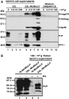
MoMuLV infection strongly enhances prion proteins release. (A) Supernatant from NIH3T3-22L (lanes 1–4), NIH3T3-22L-MoMuLV (lanes 5–8) and free complete medium as negative control (lanes 9–12) were submitted to differential centrifugation. Lanes 1, 5 and 9: 3000 g for 5 min; lanes 2, 6 and 10: 4500 g for 5 min; lanes 3, 7 and 11: 10 000 g for 30 min; and lanes 4, 8 and 12: 100 000 g for 1 h. The pellets were analyzed by Western blotting using the anti-Envgp70, anti-CAp30, anti-PrP and anti-EF1α antibodies. (B) To determine the presence of PrPSc in the 100K pellet from NIH3T3-22L-MoMuLV cells, the pellets from NIH3T3-N-MoMuLV (negative control, lane 1) and NIH3T3-22L-MoMuLV (lane 2) were treated with PK before immunoblotting with anti-PrP (lanes 3 and 4).
Figure 5
Figure 5
MoMuLV infection strongly enhances the release of prion infectivity. (A) PrPSc released is associated with MoMuLV virions and exosomes. The 100K pellets from NIH3T3-22L-MoMuLV cells were analyzed by IEM for PrP (PAG15), CAp30/Pr65Gag (PAG10) or EF1α (PAG10) after treatment by 3 M guanidium isothiocyanate (5 min). Note the presence of PrP labeling on typical dense viral particle structures (see arrows layers a1–a3) and on light spherical structures corresponding to exosomes (see black arrowheads layers a1 and a3). Single and double IEM for CAp30/Pr65Gag, PrP and EF1α, respectively, on permeabilized virions/exosomes preparation. Layers a4–a6: double IEM for CAp30/Pr65Gag and PrP on MoMuLV virions. Layer a7: EF1α labeling on exosomes (white arrowhead) and not on virions (see white arrows). Layer a9: control (Cont.) irrelevant antibody. Scale bar is 100 nm. (B) Coculture assay. The NIH3T3-N cells (target cells) were grown on the bottom surface of a six-well plate and the NIH3T3-N, NIH3T3-N-MoMuLV, NIH3T3-22L and NIH3T3-22L-MoMuLV on the surface of an insert of 0.4 μm pore size. Coculture was carried out over 4 days and target cells were passaged over 4 weeks (seven passages). Transmission of MoMuLV from the insert to the well was controlled by RT assay. Transmission of PrPSc from the insert cells to the well cells was analyzed by cell blotting in the presence or absence of PK treatment. Immunoblotting was carried out using anti-PrP.
Figure 6
Figure 6
MoMuLV anti-Env antibodies immunoprecipitate prion infectivity. (A) Experimental strategy. Magnetic beads coated with MoMuLV anti-Env (conditions 2 and 4) or with SIV anti-Vpx (as irrelevant antibody; conditions 1 and 3) were used for the immunoprecipitation experiments on NIH3T3-22L (conditions 1 and 2) and NIH3T3-22L-MoMuLV (conditions 3 and 4) supernatant. After extensive washing, RT activity was only detected on beads from condition 4 (open circle). Beads were put in contact with NIH3T3-N cells over 4 days. (B) Visualization of cell culture by direct light microscopy revealed that beads from condition 4 were still attached to the cell surface after extensive washing. (C) Cells were passaged over 6 weeks until the complete loss of beads from the culture. PK-resistant PrP was detected by cell blotting experiment in the presence or absence of PK treatment. As controls of PK treatment, NIH3T3-N and scrapie NIH3T3-22L cells were used (left panel).
Figure 7
Figure 7
MoMuLV Gag as a key factor involved in PrP release. (A) Western blotting analysis of NIH3T3-22L cellular lysate (15 μg) from cells transfected with MoMuLV GagPol, Env and GagPol+Env plasmids and probed with anti-Env, −CAp30 and −PrP antibodies. Lane 1: non-transfected NIH3T3-22L (Ct); lane 2: GagPol; lane 3: Env; and lane 4: GagPol+Env. (B) Detection of RT activity. After 3 days transfection, release of VLPs and virions was monitored by measuring RT activity in the cell supernatant. (C) GagPol release enhances PrP release. Cell supernatants were centrifuged and the 100K pellet was analyzed by Western blotting using anti-CAP30 and anti-PrP antibodies. Lane 1: 100K pellet GagPol-transfected cells; lane 2: 100K pellet Env-transfected cells; and lane 3: 100K pellet GagPol+Env-transfected cells.
Figure 8
Figure 8
PrP release correlates with MoMuLV release. (A) Structure of the MoMuLV genome and of Gag mutants affecting virus release. The gag, pol and env ORF encode, respectively, the Gag precursor composed of the matrix (MAp15), p12, capsid (CAp30) and nucleocapsid (NCp10) domains; the pol-encoded enzymes and the Envelope glycoprotein. ORFs are flanked by LTR. WT is the wild-type MoMuLV-Gag, Δp12 is Gag deleted for the p12 domain, ΔDPPPY is deleted for the DPPPY motif (L-domain) and ΔNC(16–23) is a mutant in the nucleocapsid deleted for amino acids 16–23. (B) Western blotting analyses of cell lysate (15 μg/lane) from NIH3T3-22L cells transfected with MoMuLV proviral genome DNAs with WT Gag (lane 1) or with mutant Gag Δp12 (lane 2) and ΔDPPPY (lane 3). Blots were probed with anti-CAp30, −PrP and −EF1α antibodies. (C, E) Virus production was monitored by measuring the RT activity in the supernatant. Ct corresponds to the supernatant of non-transfected NIH3T3-22L cells. (D, F) Release of PrP by MoMuLV-expressing cells. The 100K pellet containing virions and exosomes released were analyzed by Western blotting using anti-CAp30, −PrP and −EF1α antibodies. Lane 1 (D, F): MoMuLV WT; lane 2 (D): MoMuLV Gag-mutant-Δp12; lane 3 (D): mutant-ΔDPPPY; and lane 2 (F): mutant-ΔNC(16–23).

Similar articles

Cited by

References

    1. Adachi A, Gendelman HE, Koenig S, Folks T, Willey R, Rabson A, Martin MA (1986) Production of acquired immunodeficiency syndrome-associated retrovirus in human and nonhuman cells transfected with an infectious molecular clone. J Virol 59: 284–291 - PMC - PubMed
    1. Akowitz A, Sklaviadis T, Manuelidis L (1994) Endogenous viral complexes with long RNA cosediment with the agent of Creutzfeldt–Jakob disease. Nucleic Acids Res 22: 1101–1107 - PMC - PubMed
    1. Baron GS, Wehrly K, Dorward DW, Chesebro B, Caughey B (2002) Conversion of raft associated prion protein to the protease-resistant state requires insertion of PrP-res (PrP(Sc)) into contiguous membranes. EMBO J 21: 1031–1040 - PMC - PubMed
    1. Basyuk E, Galli T, Mougel M, Blanchard JM, Sitbon M, Bertrand E (2003) Retroviral genomic RNAs are transported to the plasma membrane by endosomal vesicles. Dev Cell 5: 161–174 - PubMed
    1. Bosque PJ, Prusiner SB (2000) Cultured cell sublines highly susceptible to prion infection. J Virol 74: 4377–4386 - PMC - PubMed

Publication types

MeSH terms