. 2006 Jun 21;25(12):2698-709.
doi: 10.1038/sj.emboj.7601157. Epub 2006 May 25.

Coordinated action of NSF and PKC regulates GABAB receptor signaling efficacy

Affiliations

Coordinated action of NSF and PKC regulates GABAB receptor signaling efficacy

Stéphanie M Pontier et al. EMBO J. .

Abstract

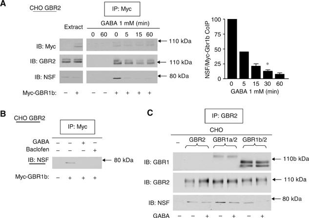
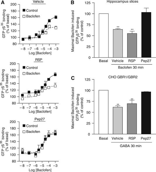
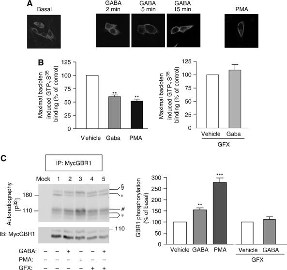
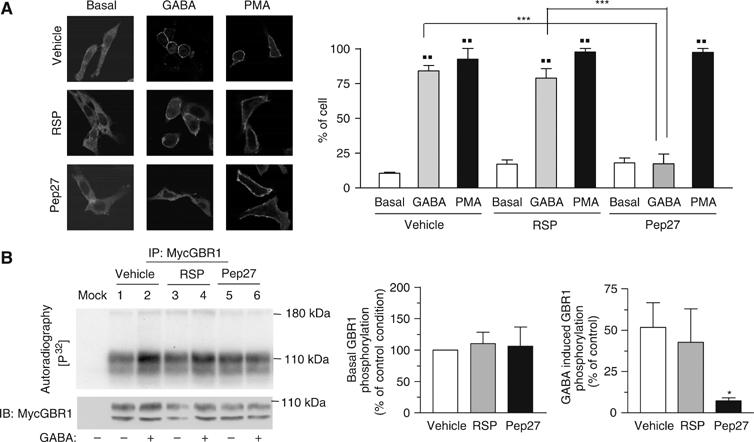
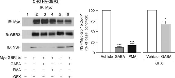
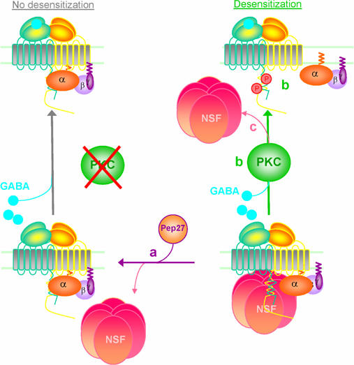
The obligatory heterodimerization of the GABAB receptor (GBR) raises fundamental questions about molecular mechanisms controlling its signaling efficacy. Here, we show that NEM sensitive fusion (NSF) protein interacts directly with the GBR heterodimer both in rat brain synaptosomes and in CHO cells, forming a ternary complex that can be regulated by agonist stimulation. Inhibition of NSF binding with a peptide derived from GBR2 (TAT-Pep-27) did not affect basal signaling activity but almost completely abolished agonist-promoted GBR desensitization in both CHO cells and hippocampal slices. Taken with the role of PKC in the desensitization process, our observation that TAT-Pep-27 prevented both agonist-promoted recruitment of PKC and receptor phosphorylation suggests that NSF is a priming factor required for GBR desensitization. Given that GBR desensitization does not involve receptor internalization, the NSF/PKC coordinated action revealed herein suggests that NSF can regulate GPCR signalling efficacy independently of its role in membrane trafficking. The functional interaction between three bona fide regulators of neurotransmitter release, such as GBR, NSF and PKC, could shed new light on the modulation of presynaptic GBR action.


Figures

Figure 1
Figure 1
Interaction between NSF and GBR c-termini assessed in yeast two-hybrid system. GBR c-termini (see schematic representation) were used as bait in a yeast two-hybrid screen testing a human brain cDNA library. NSF was one of the positive clones obtained with the GBR2 c-terminus. To delineate the GBR2 minimal region of interaction with NSF, two-hybrid assay was performed between the indicated GBR2 c-tail mutants (see table) and NSF. β-Gal activity is determined (+/−) for each construct.
Figure 2
Figure 2
Influence of the ATPase state of NSF on its direct interaction with GBR c-termini. For all experiments, purified His6-NSF was incubated with GST or GST-hybrid proteins and bound NSF was revealed by Western blot analysis. In (A) GST pull-down assay was performed with increasing amount of His6-NSF and GST or GST-GBR2ct in the presence of ATPγS and MgCl2. The graphic representation of NSF binding to the c-terminus of GBR2 is shown on the right. (B) The binding of 50 nM NSF to GST or GST-GBR2ct was assessed under the indicated nucleotide and ionic conditions. (C) GST-Pep27, GST-Pep2m or GST-RSP were incubated with 50 nM NSF in the presence of ATP, MgCl2 and EDTA. (D) GST-GBR1ct or GST-GBR2ct were incubated with 50 nM NSF in the presence of ATP, MgCl2 and EDTA. In all cases, results are representative of two to three independent experiments.
Figure 3
Figure 3
Association of NSF with the functional GBR heterodimer. Immunoprecipitations were performed on lysates from control CHO (Mock) or CHO cells stably expressing myc-GBR1b (GBR1, A), HA-GBR2 (GBR2, B), using either 9E10 anti-myc, anti-GBR2, or mouse anti-NSF antibodies. The presence of endogenous NSF or of receptors was confirmed by Western blot analysis using either 3F10 anti-myc, anti-GBR2, or rabbit anti-NSF antibodies. (C) CHO cells were transiently transfected with indicated plasmids. Cell lysates were then immunoprecipitated with 9E10 anti-myc antibody. For surface immunoprecipitation, attached cells were labeled with 9E10 anti-myc antibody before cell lysis. (D) GST-TAT hybrid proteins fused to the indicated peptide (see schematic representation) were used in order to inhibit the NSF/heterodimer interaction in CHO cells stably expressing or not expressing (Mock) myc-GBR1b/HA-GBR2. NSF/receptor complexes were immunoprecipitated with an anti-GBR2 antibody.
Figure 4
Figure 4
NSF co-localizes with both GBR subunits at the plasma membrane. (A) CHO cells stably expressing myc-GBR1b/HA-GBR2 were used. Myc-GBR1b, GBR2 and NSF were respectively labeled for immunofluorescence experiments with the secondary antibodies coupled to Alexa633 (Blue) (a), Oregon green (b) and Texas red (c) fluorophore respectively. GBR1 and GBR2 were surface labeled before cell fixation and permeabilization was then carried out in order to mark NSF. Overlay panels (d, e and f) correspond to the superposition of the a-b, a-c and b-c images, respectively. (B) GBR2 (green, a, d and g) and NSF (red, b, e and h) co-localizations (c, f and i) were determined in primary cultures of cortical neurons. Arrows indicate synaptic structures where both proteins are co-localized. White bars represent the scale of the image (20 μm).
Figure 5
Figure 5
GABA stimulation disrupts the NSF/GBR complex. (A) CHO cells stably expressing HA-GBR2 were transfected or not transfected with myc-GBR1b and were treated with GABA for the indicated times. Following immunoprecipitation with 9E10 anti-myc, the amount of precipitated NSF was revealed by Western blot and quantified (bar graph). These results represent the means±s.e.m. of three to five experiments performed independently. (B) Cells were treated either with 1 mM GABA or with 0.1 mM baclofen for 30 min. In (C) control CHO cells (−) or CHO cells stably expressing HA-GBR2 alone (GBR2), GBR1a/GBR2 or myc-GBR1b/HA-GBR2 (GBR1b/GBR2) were used and treated or not treated with GABA 1 mM for 30 min. GBR2 was immunoprecipitated and the presence of endogenous NSF and receptors revealed by Western blot.
Figure 6
Figure 6
Preventing NSF binding preserves GBR activity following GABA prestimulation. (A) Hippocampus slices were treated for 1 h with vehicle or the indicated TAT-peptide and then with 0.1 mM baclofen (empty square) or not (full square) for an additional 30 min. Curves represent the specific GTPγS binding obtained for increasing amount of baclofen. (B) Bar graph representation of the maximal baclofen induced GTPγS binding obtained from the curves in (A). (C) Similar experiments were performed in CHO cells stably expressing GBR1a and GBR2. Cells were stimulated for 30 min with 1 mM GABA.
Figure 7
Figure 7
PKC is implicated in the desensitization process of the GBR. (A) CHO cells stably expressing GBR1a/GBR2 were transiently transfected with GFP-PKCα construct. Cells were treated as indicated before their fixation and PKC localization was visualized by confocal microscopy. These results are representative of two independent experiments. (B) Maximal baclofen-stimulated [35S]GTPγS binding was measured in membranes derived from CHO cells stably expressing GBR1a/GBR2. The effect of agonist prestimulation on receptor activity was measured in the absence or presence of 0.5 μM GFX and compared to the one induced by PMA. These results represent the mean±s.e.m. of three independent experiments. (C) CHO cells stably expressing myc-GBR1b/HA-GBR2 were labeled with [32P]Pi. GBR1 was immunoprecipitated (IP, 9E10 anti-myc) and the level of phosphorylation analyzed by autoradiography. Bands corresponding to the GBR1 monomer (mature and immature species) and homodimer are indicated by (*), GBR2 monomer by (#) and GBR1/GBR2 heterodimer by (§). Results were normalized by quantifying the amount of precipitated receptor in each condition (IB). Histograms represent the mean±s.e.m. of five independent experiments.
Figure 8
Figure 8
GBR/NSF interaction is essential to PKC recruitment and GBR phosphorylation upon agonist stimulation. CHO cells stably expressing GBR1a/GBR2 were transiently transfected with GFP-PKC construct. (A) Cells were treated as indicated before their fixation and PKC localization was visualized by confocal microscopy. The histogram represents the percentage of cells displaying membrane PKC recruitment in each treatment condition and is the result of two independent experiences. In each experience, an average of 30 cells was considered per condition. (B) CHO cells stably expressing myc-GBR1b/HA-GBR2 were labeled with [32P]Pi and treated as indicated. GBR1 was immunoprecipitated (9E10 anti-myc, IP) and phosphorylation signal was analyzed by autoradiography. Histograms represent the mean±s.e.m. of four independent experiments.
Figure 9
Figure 9
PKC modulates the NSF/GABAB receptor interaction. Cells stably expressing HA-GBR2 and transiently transfected or not transfected with myc-GBR1b were used. Following treatments to modulate receptor and/or PKC activities, GBR1b was immunoprecipitated (9E10 anti-myc antibody) and the presence of endogenous NSF was revealed by western blot (IB) and quantified (bar graph). These results represent the mean±s.e.m. of three independent experiments.
Figure 10
Figure 10
Coordinated action of NSF and PKC regulates GABAB receptor signaling efficacy. (a) In the presence of TAT-Pep27, NSF dissociates from GBR and agonist-stimulation fails to promote desensitization. (b) Preassociation of NSF, however, primes the receptor such that agonist stimulation results in the recruitment of PKC and the ensuing phosphorylation of GBR leading to agonist-promoted desensitization. (c) PKC terminates the regulatory process by favoring NSF dissociation from the heterodimer.

References

    1. Barclay JW, Craig TJ, Fisher RJ, Ciufo LF, Evans GJ, Morgan A, Burgoyne RD (2003) Phosphorylation of Munc18 by protein kinase C regulates the kinetics of exocytosis. J Biol Chem 278: 10538–10545 - PubMed
    1. Becher A, Green A, Ige AO, Wise A, White JH, McIlhinney RA (2004) Ectopically expressed gamma-aminobutyric acid receptor B is functionally down-regulated in isolated lipid raft-enriched membranes. Biochem Biophys Res Commun 321: 981–987 - PubMed
    1. Becker-Hapak M, McAllister SS, Dowdy SF (2001) TAT-mediated protein transduction into mammalian cells. Methods 24: 247–256 - PubMed
    1. Blumberg PM (1991) Complexities of the protein kinase C pathway. Mol Carcinog 4: 339–344 - PubMed
    1. Bomberger JM, Parameswaran N, Hall CS, Aiyar N, Spielman WS (2005) Novel function for receptor activity-modifying proteins (RAMPs) in post-endocytic receptor trafficking. J Biol Chem 280: 9297–9307 - PubMed

Publication types

MeSH terms