. 2006 Jun 7;25(11):2623-33.
doi: 10.1038/sj.emboj.7601146. Epub 2006 May 25.

T-cell tolerance or function is determined by combinatorial costimulatory signals

Affiliations

T-cell tolerance or function is determined by combinatorial costimulatory signals

Roza Nurieva et al. EMBO J. .

Abstract

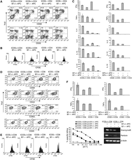
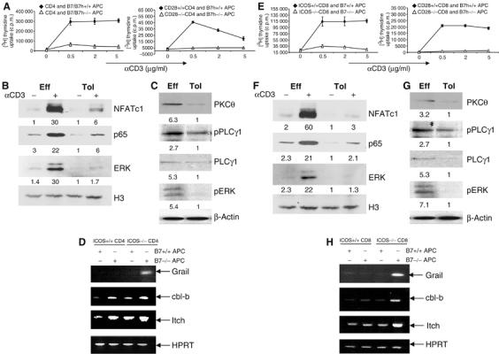
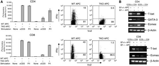
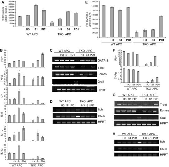
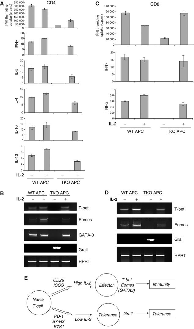
Activated in immune responses, T lymphocytes differentiate into effector cells with potent immune function. CD28 is the most prominent costimulatory receptor for T-cell activation. However, absence of CD28 costimulation did not completely impair effector function of CD4 or CD8 T cells. Moreover, increasing number of costimulatory molecules are recently found on antigen-presenting cells to regulate T-cell activation. To understand the molecular mechanisms that determine T-cell function or tolerance, we have collectively examined the roles of positive and negative costimulatory molecules. Antigen-specific naïve CD4 and CD8 T cells, only when activated in the absence of both CD28 and ICOS pathways, were completely impaired in effector function. These tolerant T cells not only were anergic with profound defects in TcR signal transduction but also completely lacked expression of effector-specific transcription factors. T-cell tolerance induction in this system requires the action by negative costimulatory molecules; T-cell proliferation and function was partially restored by inhibiting PD-1, B7-H3 or B7S1. This work demonstrates that T-cell function or tolerance is controlled by costimulatory signals.


Figures

Figure 1
Figure 1
Defective CD4 and CD8 T-cell activation in vitro in the absence of ICOS and B7 costimualation. (A) CD4+ T cells purified from ICOS +/+ or ICOS−/− OT-II mice were treated with Ova peptide in the presence of irradiated B7+/+ or B7−/− APC for 2 days, and stained with antibodies to Vα2 (PE) and CD69 or CD25 (FITC). (B) CFSE-labeled ICOS +/+ or ICOS−/− OT-II cells were activated as in (A) for 3 days. Cells were stained with Vα2-PE antibody and analyzed by FACS. Histograms represent CFSE intensity of Vα2+ populations. (C) ICOS +/+ or ICOS−/− OT-II cells were activated with Ova peptide and irradiated B7+/+ or B7−/− APC for 4 days. CD28 +/+ or CD28−/− CD4+ cells from TEa mice were treated with Ea peptide in the presence of B7h+/+ or B7h−/− APC. After 4 days, differentiated T cells were restimulated with anti-CD3 for 24 h, and effector cytokine production was measured by ELISA. All cytokines are at ng/ml. (D) CD8+ T cells from ICOS+/+ or ICOS−/− OT-I mice were stimulated with SIINFEKL peptide and irradiated B7+/+ or B7−/− APC for 2 days. Cells were stained with antibodies to Vα2 (PE) and the activation markers CD69, CD25, CD62L or CD44 (FITC). (E) CFSE-labeled ICOS+/+ and ICOS−/− OT-I cells were stimulated as in (D). CFSE division was analyzed by FACS on day 3 after T-cell activation. Cells were stained with anti-Vα2 antibodies, and analyzed by FACS. Histograms represent CFSE intensity of Vα2+ populations. (F) ICOS+/+ or ICOS −/− CD8 OT-I cells were treated with SIINFEKL peptide, and CD28+/+ or CD28 −/− CD8 2C cells with SIY peptide for 7 days in the presence of irradiated B7+/+ or B7−/− APC. Differentiated T cells were then restimulated with plate-bound anti-CD3 for 24 h and effector cytokine production was measured by ELISA. All parameters in Y-axis are ng/ml. For the CTL assay, OT-I T cells differentiated as above were incubated for 6 h with SIINFEKL peptide-pulsed EL-4 syngeneic targets. Specific lysis was calculated and shown. Differentiated OT-I T cells were analyzed for granzime B, perforin and β-actin mRNA expression by RT–PCR.
Figure 2
Figure 2
Signaling defects in T cells activated in the absence of both CD28 and ICOS costimulation. (A) CD4 OT-II cells were treated with Ova peptide in the presence of irradiated wild-type or B7−/−B7h−/− APC. CD28+/+ or CD28−/− TEa cells were treated with Ea peptide in the presence of irradiated B7h+/+ or B7h −/− APC. After 4 days, T cells were restimulated with different concentrations of anti-CD3. Proliferation was assayed 24 h after treatment by adding [3H]thymidine to the culture for the last 8 h. (B) Effector (Eff) and tolerant (Tol) OT-II cells were generated as described in (A). These cells were restimulated with plate-bound anti-CD3 for 4 h. Nuclear extracts prepared from unrestimulated and restimulated with anti-CD3 cells were probed with ERK, NFATc1 and NFκB p65 antibodies. Histon 3 (H3) was used as loading control. Relative Western blotting signals were inducted. (C) Eff and Tol CD4 cells were restimulated were restimulated with anti-CD3 for 30 min and cytoplasmic fractions were probed with PKCθ and PLCγ1 antibodies and phosphospecific antibodies against PLCγ1 and ERK. β-Actin was used as loading control. Relative Western blotting signals were inducted. (D) Grail, cbl-b, Itch and HPRT mRNA expression in restimulated effector and tolerant CD4 T cells (as in B) was analyzed by RT–PCR. (E) ICOS+/+ or ICOS −/− OT-I cells were activated with SIINFEKL peptide plus irradiated B7+/+ or B7−/− APC. CD8 cells from CD28+/+ or CD28 −/− 2C mice were treated with SIY peptide in the presence of irradiated B7h+/+ or B7h−/− APC. After 4 days, T cells were restimulated with different concentrations of plate-bound anti-CD3. Proliferation was assayed 24 h after restimulation by adding [3H]thymidine to the culture for the last 8 h. (F) Eff and Tol CD8 cells were generated as described in Figure 1F. These cells were restimulated with plate-bound anti-CD3 for 4 h. Nuclear extracts prepared from nonrestimulated and restimulated cells were probed with ERK, NFATc1 and NFκB p65 antibodies. H3 was used as loading control. Relative Western blotting signals were inducted. (G) Eff and Tol CD8 cells were restimulated with anti-CD3 for 30 min and cytoplasmic fractions were probed with PKCθ and PLCγ1 antibodies and phosphospecific antibodies against PLCγ1 and ERK. β-Actin was used as loading control. Relative Western blotting signals were inducted. (H) Grail, cbl-b, Itch and HPRT mRNA expression in restimulated effector and tolerant CD8 T cells (as in F) was analyzed by RT–PCR.
Figure 3
Figure 3
Effector defects in T cells activated in the absence of both CD28 and ICOS costimulation. (A) CD4 OT-II cells or CD8 OT-I cells were treated with Ova peptide or SIINFEKL peptide in the presence of irradiated wild-type or B7−/−B7h−/− APC. After 4 days, T cells were restimulated with anti-CD3 (5 μg/ml) or PMA/Ionomycin. Proliferation was assayed 24 h after restimulation by adding [3H]thymidine to the culture for the last 8 h. For cytokine measurement, CD4 OT-II cells or CD8 OT-I cells were treated as described above. After 5 days, T cells were restimulated with PMA/Ionomycin for 5 h. Intracellular staining of IFNγ was performed on gated Vα2+ T cells, and then analyzed by FACS. (B). T-bet, GATA3, Eomes and β-actin mRNA expression in effector and tolerant CD4 T cells (as in Figure 1C) was analyzed by RT–PCR. Effector and tolerant CD8+ T cells (from 1F) were analyzed for T-bet, Eomes and β-actin mRNA expression by RT–PCR.
Figure 4
Figure 4
Negative costimulatory molecules regulate tolerance induction of CD4 and CD8 T cells. (A) OT-II cells were treated with Ova peptide in the presence of irradiated wild-type (WT) or B7−/−B7h−/− (TKO) APC with a control rat IgG or a blocking antibody to B7S1 (S1) or B7H3 (H3) or PD1. At 4 days after activation, T cells were restimulated with plate-bound anti-CD3. Proliferation was assayed 24 h after treatment by adding [3H]thymidine to the culture for the last 8 h. (B) CD4 cells activated as in (A) were restimulated with plate-bound anti-CD3 for 24 h and cytokine production was measured by ELISA. All parameters indicated in Y-axis are at ng/ml. (C) Differentiated OT-II T cells were analyzed for GATA-3, T-bet, Eomes, Grail and HPRT mRNA expression by RT–PCR. (D) Differentiated OT-II T cells were analyzed for Itch, Cbl-b and HPRT mRNA expression by RT–PCR. (E) OT-I cells were treated with SIINFEKL peptide in the presence of irradiated WT or B7−/−B7h−/− (TKO) APC with a control rat IgG or a blocking antibody to B7S1 (S1) or B7H3 (H3) or PD1. At 6–7 days after activation, T cells were restimulated with plate-bound anti-CD3. Proliferation was assayed 24 h after treatment by adding [3H]thymidine to the culture for the last 8 h. (F) CD8 cells activated as in (E) were restimulated with plate-bound anti-CD3 for 24 h and cytokine production was measured by ELISA. All parameters indicated in Y-axis are at ng/ml. (G) Differentiated OT-I T cells were analyzed for T-bet, Eomes, Grail and HPRT mRNA expression by RT–PCR. (H) Differentiated OT-I T cells were analyzed for Itch, Cbl-b and HPRT mRNA expression by RT–PCR.
Figure 5
Figure 5
IL-2 regulates T-cell tolerance and immunity. (A–D) CD4 OT-II cells (A, B) or CD8 OT-I cells (C, D) were treated with Ova peptide or SIINFEKL peptide, respectively, in the presence of irradiated wild-type (WT) or B7−/−B7h−/− (TKO) APC with or without IL-2. After 4 days for CD4 (A) and 6–7 days for CD8 cells (C), T cells were restimulated with plate-bound α-CD3 to measure T-cell proliferation and effector cytokine (IFN-γ, IL-4, -5, -10, -13) production. Activated OT-II (B) or OT-I (D) cells were analyzed for T-bet, Eomes, Grail and HPRT mRNA expression by RT–PCR. (E) T-cell immunity or tolerance is regulated by a combinatorial costimulation signal. At resting states, when T cells encounter APC, negative costimulatory molecules PD-1, B7-H3 and B7S1 function to inactivate T cells. During infection, CD28 and ICOS ligands are upregulated on APC, which drives T cells to differentiate into effector T cells.

References

    1. Agata Y, Kawasaki A, Nishimura H, Ishida Y, Tsubata T, Yagita H, Honjo T (1996) Expression of the PD-1 antigen on the surface of stimulated mouse T and B lymphocytes. Int Immunol 8: 765–772 - PubMed
    1. Anandasabapathy N, Ford GS, Bloom D, Holness C, Paragas V, Seroogy C, Skrenta H, Hollenhorst M, Fathman CG, Soares L (2003) GRAIL: an E3 ubiquitin ligase that inhibits cytokine gene transcription is expressed in anergic CD4+ T cells. Immunity 18: 535–547 - PubMed
    1. Andris F, Denanglaire S, de Mattia F, Urbain J, Leo O (2004) Naive T cells are resistant to anergy induction by anti-CD3 antibodies. J Immunol 173: 3201–3208 - PubMed
    1. Barber DL, Wherry EJ, Masopust D, Zhu B, Allison JP, Sharpe AH, Freeman GJ, Ahmed R (2006) Restoring function in exhausted CD8T cells during chronic viral infection. Nature 439: 682–687 - PubMed
    1. Bertram EM, Tafuri A, Shahinian A, Chan VS, Hunziker L, Recher M, Ohashi PS, Mak TW, Watts TH (2002) Role of ICOS versus CD28 in antiviral immunity. Eur J Immunol 32: 3376–3385 - PubMed

Publication types

MeSH terms