. 2006 Jun 7;25(11):2539-50.
doi: 10.1038/sj.emboj.7601140. Epub 2006 May 25.

How initiation factors tune the rate of initiation of protein synthesis in bacteria

Affiliations

How initiation factors tune the rate of initiation of protein synthesis in bacteria

Ayman Antoun et al. EMBO J. .

Abstract

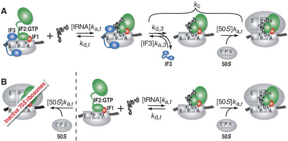
The kinetics of initiator transfer RNA (tRNA) interaction with the messenger RNA (mRNA)-programmed 30S subunit and the rate of 50S subunit docking to the 30S preinitiation complex were measured for different combinations of initiation factors in a cell-free Escherichia coli system for protein synthesis with components of high purity. The major results are summarized by a Michaelis-Menten scheme for initiation. All three initiation factors are required for maximal efficiency (kcat/KM) of initiation and for maximal in vivo rate of initiation at normal concentration of initiator tRNA. Spontaneous release of IF3 from the 30S preinitiation complex is required for subunit docking. The presence of initiator tRNA on the 30S subunit greatly increases the rate of 70S ribosome formation by increasing the rate of IF3 dissociation from the 30S subunit and the rate of 50S subunit docking to the IF3-free 30S preinitiation complex. The reasons why IF1 and IF3 are essential in E. coli are discussed in the light of the present observations.


Figures

Figure 1
Figure 1
Kinetic scheme for initiation of protein synthesis. (A) Binding of fMet-tRNAfMet to mRNA-programmed 30S subunits equipped with three initiation factors destabilizes IF3 binding. IF3 dissociates then with the rate kd,3 after which it can rebind with the rate ka,3[IF3]. Alternatively, a 50S subunit can dock to an IF3-free 30S preinitiation complex with the rate ka,r[50S]. (B) In the absence of IF3, both 50S subunits and fMet-tRNAfMet can bind with comparable rates to the mRNA-programmed 30S subunits. Binding of 50S subunits gives rise to an empty 70S ribosome inactive in initiation, whereas fMet-tRNAfMet binding results in a subsequent fast docking of 50S subunits and formation of active 70S initiation complexes.
Figure 2
Figure 2
Effect of initiation factors on fMet-tRNAfMet binding to 30S ribosomal subunits. (A) [3H]fMet-tRNAfMet association to mRNA-programmed 30S subunits was measured by NC filtration technique in the absence of initiation factors or with only IF1 present. Insert: dependence of fMet-tRNAfMet association rate on the concentration of fMet-tRNAfMet in the absence of initiation factors. (B) [3H]fMet-tRNAfMet dissociation from 30S preinitiation complexes assembled with different combinations of IF1 and IF2 was measured by NC filtration in the presence of a 10-fold excess of nonlabeled chasing fMet-tRNAfMet. (C) The same as in (B) except that IF3 was also present in the 30S preinitiation complex. (D) Dependence of the 30S·mRNA·IF3·fMet-tRNAfMet complex formation on the concentration of fMet-tRNAfMet in the presence or absence of IF1 measured by NC filtration. (E) Kinetics of fMet-tRNAfMet binding to 30S·mRNA·IF2 or to 30S·mRNA·IF1·IF2 complexes measured by light scattering. (F) The same as in (E) but in the presence of IF3.
Figure 3
Figure 3
Effect of initiation factors on the association of ribosomal subunits. (A) 50S subunit docking to mRNA-programmed 30S complexes containing different combinations of IF1 and IF2 in the absence of fMet-tRNAfMet measured by light scattering. (B) The same as in (A) but all 30S complexes contained IF3. The gray curves show dissociation of the corresponding 70S complexes assembled without IF3 after IF3 addition. (C) 50S subunit docking to 30S complexes containing fMet-tRNAfMet and different combinations of IF1 and IF2 measured by light scattering. Insert: extended time range. (D) The same as in (C) but 30S complexes contained IF3. Insert: extended time range. (E) Rate of 50S subunit docking to 30S preinitiation complexes monitored as the rate of dipeptide formation in a quench-flow experiment. One syringe of the quench-flow instrument contained 50S subunits and ternary complexes, whereas the other contained 30S preinitiation complexes assembled with all three initiation factors, or in the absence of either IF1 or IF3. The time course of the dipeptide formation reaction (peptidyl transfer) with preformed 70S initiation complexes is shown for comparison.
Figure 4
Figure 4
Formation of 70S ribosomes requires IF3 dissociation from 30S subunits. (A) Dependence of the rate of the 50S subunit docking to the 30S·mRNA·IF2·fMet-tRNAfMet complex or to the same complex containing IF1 on the concentration of 50S subunits. Insert: the rate of 70S formation from empty 30S and 50S subunits plotted versus the concentration of 50S subunits. Error bars smaller than symbol size are not visible. (B) The same as in (A) but 30S complexes also contained IF3. (C) The rate dependences in (B) replotted as reciprocal of the rate versus the reciprocal of the concentration of 50S subunits. Error bars smaller than symbol size are not visible. (D) The reciprocal of the rate of the 50S subunit docking to the 30S·mRNA·IF3·IF2·fMet-tRNAfMet complex or to the same complex containing IF1 plotted versus the concentration of IF3 in the reaction mixture. Error bars smaller than symbol size are not visible.
Figure 5
Figure 5
IF3 prevents the formation of abortive 70S initiation complexes. (A) Competition between IF3 and the 50S subunit for binding to the 30S·mRNA·IF2·fMet-tRNAfMet complex; 50S subunits were added to the 30S complexes or to the same complexes containing IF3. Alternatively, a mixture containing 50S subunits and IF3 was added to the 30S complexes. (B) Kinetics of the formation of 70S ribosomes from 30S·mRNA·IF2 complexes containing different combinations of IF1 and IF3 after addition of 50S subunits together with fMet-tRNAfMet. Alternatively, 70S ribosomes were formed by mixing 30S·IF1·IF2·IF3 complexes with a mixture containing mRNA, 50S subunits and fMet-tRNAfMet. (C) fMet-Phe dipeptide formation after the addition of 30S·mRNA·IF1·IF2 complexes to the mixture B containing EF-Tu·GTP·Phe-tRNAPhe ternary complexes, fMet-tRNAfMet and 50S subunits in 0.45 μM or 0.9 μM concentration. The reaction was also repeated with 30S·mRNA·IF1·IF2·IF3 complexes and the same mixture B containing 0.45 μM 50S subunits. (D) Reduction in the concentration of fMet-tRNAfMet containing 70S ribosomes assembled by addition of 30S·mRNA·IF2 complexes to a mixture containing increasing concentrations of 50S subunits and fMet-tRNAfMet at fixed concentration. 30S·mRNA·IF2 complexes contained different combinations of IF1 and IF3. The amount of 70S ribosomes containing [3H]fMet-tRNAfMet was measured by nitrocellulose filtration after 30 s from the start of the assembly reaction.

References

    1. Allen GS, Zavialov A, Gursky R, Ehrenberg M, Frank J (2005) The cryo-EM structure of a translation initiation complex from Escherichia coli. Cell 121: 703–712 - PubMed
    1. Antoun A, Pavlov MY, Andersson K, Tenson T, Ehrenberg M (2003) The roles of initiation factor 2 and guanosine triphosphate in initiation of protein synthesis. EMBO J 22: 5593–5601 - PMC - PubMed
    1. Antoun A, Pavlov MY, Tenson T, Ehrenberg MM (2004) Ribosome formation from subunits studied by stopped-flow and Rayleigh light scattering. Biol Proc Online 6: 35–54 - PMC - PubMed
    1. Benne R, Ebes F, Voorma HO (1973) Sequence of events in initiation of protein synthesis. Eur J Biochem 38: 265–273 - PubMed
    1. Canonaco MA, Calogero RA, Gualerzi CO (1986) Mechanism of translational initiation in prokaryotes. Evidence for a direct effect of IF2 on the activity of the 30 S ribosomal subunit. FEBS Lett 207: 198–204 - PubMed

Publication types

MeSH terms

LinkOut - more resources