. 2006 Sep 1;398(2):303-10.
doi: 10.1042/BJ20060300.

Novel transcripts of Nox1 are regulated by alternative promoters and expressed under phenotypic modulation of vascular smooth muscle cells

Affiliations

Novel transcripts of Nox1 are regulated by alternative promoters and expressed under phenotypic modulation of vascular smooth muscle cells

Noriaki Arakawa et al. Biochem J. .

Abstract

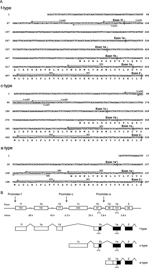
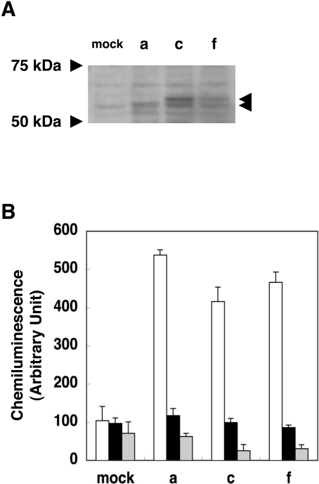
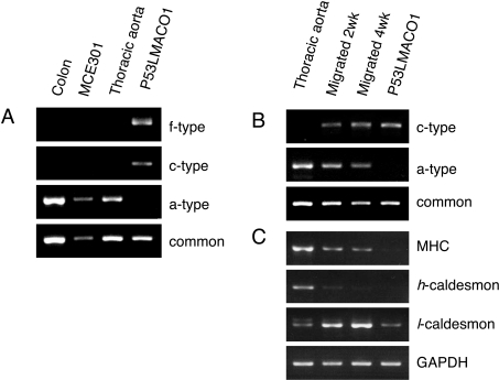
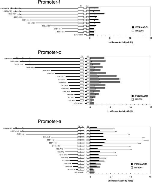
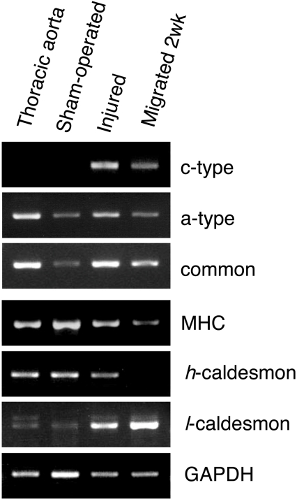
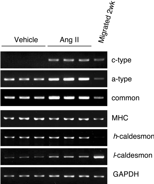
NADPH oxidase is implicated in the pathogenesis of various cardiovascular disorders. In vascular smooth muscle cells (VSMC), expression of NOX1 (NADPH oxidase 1), a catalytic subunit of NADPH oxidase, is low and is induced upon stimulation by vasoactive factors, while it is abundantly expressed in colon epithelial cells. To clarify the regulatory mechanisms underlying such cell-specific expression, the upstream regions directing transcription of the NOX1 gene were explored. In P53LMACO1 cells, a cell line originated from mouse VSMCs, two novel Nox1 mRNA species, the c- and f-type, were isolated. These transcripts contained 5'-untranslated regions that differed from the colon type mRNA (a-type) and encoded an additional N-terminal peptide of 28 amino acids. When these transcripts were fused to the c-myc tag and expressed in human embryonic kidney 293 cells, a fraction of translated proteins demonstrated the size containing the additional peptide. Proteins encoded by the c- and f-type mRNAs exhibited superoxide-producing activities equivalent to the activity of the a-type form. The a-type mRNA was expressed in the colon and in the intact aorta, whereas the c-type mRNA was detected in the primary cultured VSMCs migrated from aortic explants, in vascular tissue of a wire-injury model and in the thoracic aorta of mice infused with angiotensin II. The promoter region of the c-type mRNA exhibited transcriptional activity in P53LMACO1 cells, but not in MCE301 cells, a mouse colon epithelial cell line. These results suggest that expression of the Nox1 gene is regulated by alternative promoters and that the novel c-type transcript is induced under phenotypic modulation of VSMCs.


Figures

Figure 1
Figure 1. Structure of the novel transcript forms of the Nox1 gene
(A) 5′-Nucleotide sequences of the f-, c- and a-type Nox1 cDNA and deduced amino acid sequences at the beginning of open reading frames. Translational start sites are underlined. Primers used for 5′-RACE are indicated with arrows. (B) The exon/intron structure of the Nox1 gene and its alternative splicing pathways. Open boxes indicate exons. Numbers in the box denote the size of each exon (bp). The size of the intron is indicated under the broken lines (bp). Closed boxes show the open reading frames of the transcript.
Figure 2
Figure 2. Characterization of NOX1 proteins encoded by the novel transcript forms
(A) NOX1–c-myc fusion proteins expressed in HEK-293 cells. Cells were transfected with vectors containing the f-, c- or a-type Nox1–c-myc tag fusion cDNA. Whole cell lysates (10 μg) were separated by SDS/PAGE (8.4% gels) and the c-myc-tagged proteins were detected with the mouse monoclonal antibody as described in the Experimental section. (B) Superoxide-producing activities of NOX1 proteins encoded by the novel transcripts. HEK-293 cells transfected with pcDNA3 containing Nox1, NOXA1 and NOXO1 cDNAs were incubated for 5 min in Krebs/Hepes buffer containing 5 μM lucigenin and 100 μM NADPH in the presence or absence of 10 μM diphenyleneiodonium chloride (DPI). The chemiluminescence was measured by a luminometer and expressed as means±SE (n=4). Open bar, cells transfected with NOX1, NOXA1 and NOXO1; closed bar, NOX1 and NOXA1 and NOXO1 and DPI; grey bar, NOX1 only.
Figure 3
Figure 3. Cell-specific expression of the Nox1 transcripts
(A) Expression of the f-, c-, and a-type Nox1 transcripts in mouse tissues and cell lines. Nox1 transcripts containing different 5′-UTRs or the common sequence (common) were amplified by RT-PCR with total RNA from indicated tissues and cells, and separated on 2.5% agarose gel. (B) Expression of the c-type transcript in migrated VSMCs. VSMCs migrated from aortic explants were cultured for 2- or 4-weeks, and expression of each transcript form was analysed by RT-PCR. (C) Expression of smooth muscle-specific molecular markers. Myosin heavy chain (MHC), h-, l-caldesmon and GAPDH (glyceraldehyde-3-phosphate dehydrogenase) mRNAs were detected in the total RNA used in (B). Figures are representative of at least three independent experiments.
Figure 4
Figure 4. Analyses of the Nox1 promoters in different cell lines
(A) Schematic diagram of the promoter-luciferase fusion plasmids is shown on the left, where the 5′/3′ ends of the construct relative to the transcription initiation site are indicated. Each construct was transiently transfected into P53LMACO1 (closed bar) or MCE301 cells (opened bar). The β-galactosidase-expression vector was co-transfected as an internal control. The relative luciferase activity was denoted as the ratio to the activity of the pGL3-basic vector. Bars represent means ±SE of at least three experiments.
Figure 5
Figure 5. Expression of the c-type Nox1 transcript in injured arteries
Nox1 mRNA containing different 5′-UTRs, the common sequence (common), smooth muscle-specific molecular markers and GAPDH (glyceraldehyde-3-phosphate dehydrogenase) were detected by RT-PCR. A wire-injury was inflicted in the femoral artery of the left leg of each mouse. At 14 days after the surgery, total RNA was extracted from pooled arteries from the sham-operated right legs or the injured left legs of three mice. The thoracic aorta and migrated VSMCs cultured for 2 weeks were used as controls.
Figure 6
Figure 6. Expression of the c-type Nox1 transcript in the thoracic aorta of mice infused with angiotensin II
Nox1 mRNA containing different 5′-UTRs, the common sequence (common), smooth muscle-specific molecular markers and GAPDH (glyceraldehyde-3-phosphate dehydrogenase) were detected by RT-PCR. Total RNA was isolated from three mice infused with angiotensin II or vehicle for 7 days.

References

    1. Suh Y. A., Arnold R. S., Lassègue B., Shi J., Xu X., Sorescu D., Chung A. B., Griendling K. K., Lambeth J. D. Cell transformation by the superoxide-generating oxidase Mox1. Nature (London) 1999;401:79–82. - PubMed
    1. Bánfi B., Maturana A., Jaconi S., Arnaudeau S., Laforge T., Sinha B., Ligeti E., Demaurex N., Krause K. H. A mammalian H+ channel generated through alternative splicing of the NADPH oxidase homolog NOH-1. Science. 2000;287:138–142. - PubMed
    1. Lassègue B., Clempus R. E. Vascular NAD(P)H oxidases: specific features, expression, and regulation. Am. J. Physiol. Regul. Integr. Comp. Physiol. 2003;285:R277–R297. - PubMed
    1. Lassègue B., Sorescu D., Szöcs K., Yin Q., Akers M., Zhang Y., Grant S. L., Lambeth J. D., Griendling K. K. Novel gp91phox homologues in vascular smooth muscle cells: nox1 mediates angiotensin II-induced superoxide formation and redox-sensitive signaling pathways. Circ. Res. 2001;88:888–894. - PubMed
    1. Katsuyama M., Fan C., Yabe-Nishimura C. NADPH oxidase is involved in prostaglandin F2α-induced hypertrophy of vascular smooth muscle cells: induction of NOX1 by PGF2α. J. Biol. Chem. 2002;277:13438–13442. - PubMed

Publication types

MeSH terms

Associated data