. 2006 May 25:4:22.
doi: 10.1186/1479-5876-4-22.

Induction of antitumor immunity through xenoplacental immunization

Affiliations

Induction of antitumor immunity through xenoplacental immunization

Zhaohui Zhong et al. J Transl Med. .

Abstract

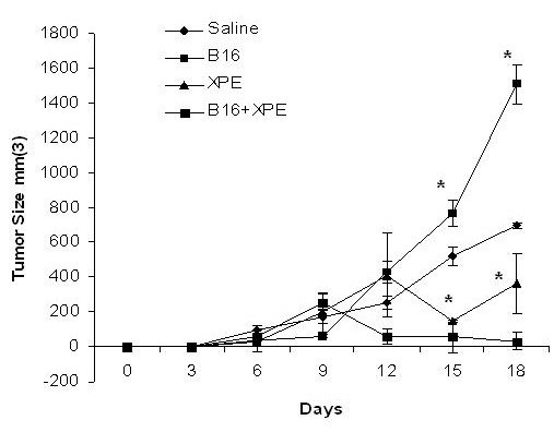
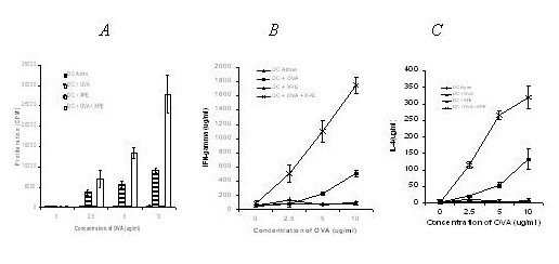
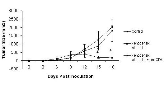
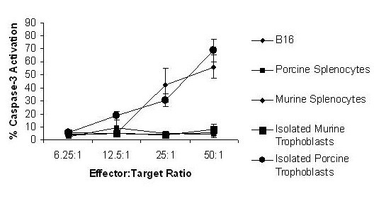
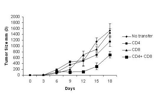
Historically cancer vaccines have yielded suboptimal clinical results. We have developed a novel strategy for eliciting antitumor immunity based upon homology between neoplastic tissue and the developing placenta. Placenta formation shares several key processes with neoplasia, namely: angiogenesis, activation of matrix metalloproteases, and active suppression of immune function. Immune responses against xenoantigens are well known to break self-tolerance. Utilizing xenogeneic placental protein extracts as a vaccine, we have successfully induced anti-tumor immunity against B16 melanoma in C57/BL6 mice, whereas control xenogeneic extracts and B16 tumor extracts where ineffective, or actually promoted tumor growth, respectively. Furthermore, dendritic cells were able to prime tumor immunity when pulsed with the placental xenoantigens. While vaccination-induced tumor regression was abolished in mice depleted of CD4 T cells, both CD4 and CD8 cells were needed to adoptively transfer immunity to naïve mice. Supporting the role of CD8 cells in controlling tumor growth are findings that only freshly isolated CD8 cells from immunized mice were capable of inducing tumor cell caspases-3 activation ex vivo. These data suggest feasibility of using xenogeneic placental preparations as a multivalent vaccine potently targeting not just tumor antigens, but processes that are essential for tumor maintenance of malignant potential.


Figures

Figure 1
Figure 1
Antitumor effects of XPE. XPE preparations were dissolved into sterile, injection-grade PBS at a concentration of 2 mg/ml, and injections of 50 uL (total mass 10 ug) were performed subcutaneously into C57/BL6 mice 7 days before tumor challenge. 5 × 10⁵ B16 murine melanoma cells were injected subcutaneously into the hind limb flank of female 6–8 week-old C57BL/6. Tumor growth was assessed every 3 days by two measurements of perpendicular diameters by a caliper, and animals were sacrificed when tumors reached a size of 1 cm in any direction. *p < 0.05, Student's T test compared to saline treated.
Figure 2
Figure 2
XPE augments immunity of B16 lysate "vaccine". XPE and B16 melanoma cell lysates were prepared as described in Figure 1 using freeze-thaw cycles. C57/BL6 mice were immunized with XPE alone or co-mixed with the B16 melanoma lysate diluted in PBS at a concentration of 2 mg/ml, and injections of 50 uL (total mass 10 ug). Tumor growth was assessed every 3 days by two measurements of perpendicular diameters by a caliper, and animals were sacrificed when tumors reached a size of 1 cm in any direction. *p < 0.05, Student's T test compared to saline treated.
Figure 3
Figure 3
Induction of anti-cancer immunity by XPE-pulsed DC. Day 7 bone marrow-derived DC were pulsed with 10 μg/ml XPE for 24 h and injected s.c. (5 × 10⁵ cells/mouse) into syngeneic C57BL/6 mice. A concurrent injection of 5 × 10⁵ B16 melanoma cells was administered. Tumor growth was assessed every 3 days by two measurements of perpendicular diameters by a caliper, and animals were sacrificed when tumors reached a size of 1 cm in any direction. *p < 0.05, Student's T test compared to saline treated.
Figure 4
Figure 4
Potentiation of immunity by XPE pulsed DC. C57/BL6 mice were injected were injected subcutaneously with 5 × 10⁵ bone marrow-derived DC pulsed with either 10 μg/ml XPE, and/or 10 μg/ml OVA for 24 hr. Mice were sacrificed 14 days post inoculation and splenocytes were cultured with increasing concentrations of OVA in vitro. Cultures were assessed for: A) Proliferation; B) IFN-γ production; and C) IL-4 production.
Figure 5
Figure 5
XPE-induced anti-tumor immunity is CD4 dependent. CD4 cell depletion was accomplished in C57/BL6 mice by intravenous injection of anti-CD4 monoclonal antibody (clone GK1.5) on day -5, day -3, day 0, day 1, day 3, and day 5 at a concentration of 150 ug/mouse. XPE was injected on day 0, which was also the timepoint of tumor injection (5 × 10⁵ B16) Tumor growth was assessed every 3 days by two measurements of perpendicular diameters by a caliper, and animals were sacrificed when tumors reached a size of 1 cm in any direction. *p < 0.05, Student's T test compared to saline treated.
Figure 6
Figure 6
XPE-induction of caspase-3 activating CD8 cells. CD8 T cells were isolated from spleens of experimental and control mice on day 8 after immunization with XPE and mixed at the indicated ratios with the indicated target cells. Caspase-3 activation was quantified as percentage of target cells positive for anti-activated caspase-3 antibody staining by flow cytometry. The results are representative of 3 independently performed experiments.
Figure 7
Figure 7
Adoptive transfer of XPE-induced immunity. Mice were immunized with XPE, challenged on day 7 with 5 × 10⁵ B16 cells, observed for an additional 18 days after which CD4+, and CD8+ cells were harvested from splenocytes. CD4 and CD8 cells were transferred alone or together at a concentration of 107 cells/mouse to naïve C57/BL6 mice intravenously at time of tumor challenge with 5 × 10⁵ B16 cells.

Similar articles

Cited by

References

    1. Regnault TR, Galan HL, Parker TA, Anthony RV. Placental development in normal and compromised pregnancies-- a review. Placenta. 2002;23 Suppl A:S119–29. doi: 10.1053/plac.2002.0792. - DOI - PubMed
    1. Curry TEJ, Osteen KG. The matrix metalloproteinase system: changes, regulation, and impact throughout the ovarian and uterine reproductive cycle. Endocr Rev. 2003;24:428–465. doi: 10.1210/er.2002-0005. - DOI - PubMed
    1. Szekeres-Bartho J. Immunological relationship between the mother and the fetus. Int Rev Immunol. 2002;21:471–495. doi: 10.1080/08830180215017. - DOI - PubMed
    1. Pijnenborg R. Implantation and immunology: maternal inflammatory and immune cellular responses to implantation and trophoblast invasion. Reprod Biomed Online. 2002;4 Suppl 3:14–17. - PubMed
    1. Bischof P, Meisser A, Campana A. Control of MMP-9 expression at the maternal-fetal interface. J Reprod Immunol. 2002;55:3–10. doi: 10.1016/S0165-0378(01)00142-5. - DOI - PubMed