Comparative Study
. 2005:1:2005.0008.
doi: 10.1038/msb4100012. Epub 2005 May 25.

Phosphotyrosine interactome of the ErbB-receptor kinase family

Affiliations
Comparative Study

Phosphotyrosine interactome of the ErbB-receptor kinase family

Waltraud X Schulze et al. Mol Syst Biol. 2005.

Abstract

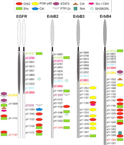
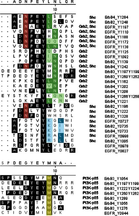
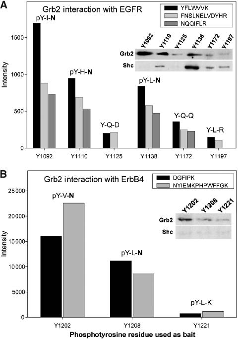
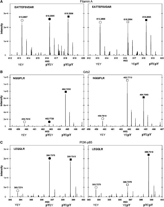
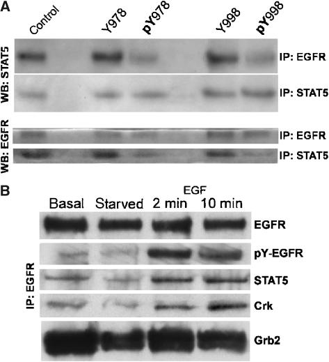
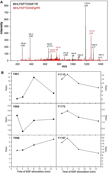
Interactions between short modified peptide motifs and modular protein domains are central events in cell signal-transduction. We determined interaction partners to all cytosolic tyrosine residues of the four members of the ErbB-receptor family in an unbiased fashion by quantitative proteomics using pull-down experiments with pairs of phosphorylated and nonphosphorylated synthetic peptides. Each receptor had characteristic preferences for interacting proteins and most interaction partners had multiple binding sites on each receptor. EGFR and ErbB4 had several docking sites for Grb2, while ErbB3 was characterized by six binding sites for PI3K. We identified STAT5 as a direct binding partner to EGFR and ErbB4 and discovered new recognition motifs for Shc and STAT5. The overall pattern of interaction partners of EGFR and ErbB4 suggests similar roles during signaling through their respective ligands. Phosphorylation kinetics of several tyrosine resides was measured by mass spectrometry and correlated with interaction partner preference. Our results demonstrate that system-wide mapping of peptide-protein interactions sites is possible, and suggest shared and unique roles of ErbB-receptor family members in downstream signaling.


Figures

Figure 1
Figure 1
Summary of systematic interaction profiling of the ErbB-receptor tyrosine family. All cytosolic residues of the ErbB-receptor family were used in pull-down assays. The interaction partners found are indicated by symbols. The kinase domain of the receptors is designated as an oval. Underlined and colored tyrosine residues mark identical sequence regions between different receptors; regions around colored residues show strong homology between receptors. Most interaction partners to tyrosine residues are found at the C-terminal end outside the kinase domain. The EGFR has multiple interaction partners, and several binding sites for Grb2. ErbB2 has few interaction partners; of them Shc is the most common. ErbB3 interacts mainly with P13-Kinase subunit p85, and ErbB4 again shows a diversity of interaction partners, also with several binding sites for Grb2. Receptors are not drawn to scale. The dimerization is indicated by EGFR dimer. Residues are labeled according to the full-length sequence of each of the receptors.
Figure 2
Figure 2
Binding motifs of the major interaction partners of the ErbB-receptor family. Consensus motifs for binding of Grb2, Shc and P13-Kinase are presented. Sequences shaded in black are common to the majority of all aligned sequences. Residues shaded in red denote essential residues for PTB-domain binding of Shc, while green shades indicate Grb2 binding sites. Residues shaded in blue indicate a motif for SH2-domain binding of Shc. Yellow residues mark essential amino acids in the binding motif for the SH2 domain of P13K. The binding motif for the PTB domain of Shc is N-P-X-pY. The residue N at position −3 is essential, whereas the residue P at position −2 can also be replaced. Besides the known Grb2-binding motif pY-X-N, five binding sites were found for Grb2, which did not contain an N in position +2 after the phosphotyrosine. A binding target for the SH2 domain of Shc was found to contain pY-[KR]-X-φ. The binding motif for P13-Kinase subunit p85 is pY-X-X-M.
Figure 3
Figure 3
Interaction strength of Grb2 binding sites. Ion intensities of extracted ion chromatograms of identical tryptic peptides of Grb2, which were identified in pull-down experiments using different phosphotyrosine containing bait peptides. The two amino acids C-terminal to the bait phosphotyrosine are indicated. (A) Ion intensities of tryptic Grb2 peptides in pull-downs with phosphotyrosine baits of EGFR. (B) Ion intensities of tryptic Grb2 peptides in pull-downs with phosphotyrosine baits of ErbB4. Insets show abundance of interaction partner by Western blot.
Figure 4
Figure 4
Interaction partners with the YEY motifs of ErbB3. Using metabolic labeling with Arg0, Arg6 and Arg10, interaction partners to Y-E-Y in all three phosphorylation combinations were determined. Results are shown for the two tyrosines around Yl 197 and Yl 199. (A) MS spectra of a tryptic peptide of Filamin A, an unspecific binding protein as indicated by the same ion intensity when precipitated by the nonphosphorylated, singly phosphorylated and doubly phosphorylated bait. (B) MS spectra of a tryptic peptide of Grb2. Grb2 binds to pY-E-pY and Y-E-pY. (C) MS spectra of a tryptic peptide of PI3-Kinase. PI3-Kinase subunit p85 binds to doubly phosphorylated peptide and pY-E-Y. No protein was found to bind specifically to the doubly phosphorylated peptides.
Figure 5
Figure 5
Co-immunoprecipitation of STAT5 and EGFR. (A) In co-immunoprecipitation experiments, EGFR was detected by Western blot (WB) in immunoprecipitations (IP) with STAT5, and STAT5 was detected in immunoprecipitations of EGFR. Co-immunoprecipitation was reduced by addition of phosphorylated peptides pY978 and pY998 of EGFR. There was no effect when these peptides were added in their nonphosphorylated form as control (Y978 and Y998). In addition, equal amounts of EGFR and STAT5 were immunoprecipitated, as shown by the IP with STAT5, WB with STAT5 and the IP with EGFR, WB with EGFR, respectively. The experiments were performed on HeLa cells growing in 10% FBS. (B) Phospho-EGFR, STAT5 and Crk after immunoprecipitation of EGFR from normal growing HeLa cells (basal), serum-starved cells for 14 h (starved) and EGF-stimulated cells for 2 and 10 min. The antibody was used for IP and WB of EGFR against codons 998-1022, while the antibody against phospho-EGFR was directed against pYl 197 of EGFR. STAT5 antibody is directed against the C-terminal domain of Stat5b, but recognized isoforms STAT5a/b, and Crk antibody is directed against the N-terminus of Crk.
Figure 6
Figure 6
Phosphorylation dynamics of EGFR. (A) MS-MS fragmentation spectrum of the tryptic peptide MHLPSPTDSNFYR and MHLPSPTDSNF[pY]R of EGFR showing phosphorylation of tyrosine 998. (B) Time course of phosphorylation at six different tyrosine residues of the EGFR upon EGF stimulation. Ratios of peak intensities after EGF stimulation versus no stimulation are shown. Tyrosine residues Y1110, Y1172 and Y1197 are autophosphorylation sites, tyrosine Y869 is phosphorylated by Src kinase, and tyrosine Y998 and Y801 are novel phosphorylation sites.

Similar articles

Cited by

References

    1. Andersen JS, Wilkinson CJ, Myoru T, Mortensen P, Nigg EA, Mann M (2003) Proteomic characterization of the human centrosome by protein correlation profiling. Nature 426: 570–574 - PubMed
    1. Birge RB, Fajardo JE, Mayer BJ, Hanafusa H (1992) Tyrosine-phosphorylated epidermal growth factor receptor and cellular p130 provide high affinity binding substrates to analyze Crk-phosphotyrosine-dependent interactions in vitro. J Biol Chem 267: 10588–10595 - PubMed
    1. Blagoev B, Kratchmarova I, Ong S-E, Nielsen M, Foster L, Mann M (2003) A proteomics strategy to elucidate functional protein-protein interactions applied to EGF signaling. Nat Biotechnol 21: 315–318 - PubMed
    1. Blagoev B, Ong S-E, Kratchmarova I, Mann M (2004) Temporal ordering of signaling networks by quantitive proteomics. Nat Biotechnol 22: 1139–1145 - PubMed
    1. Blume-Jensen P, Hunter T (2001) Oncogenic kinase signalling. Nature 411: 355–365 - PubMed

Publication types

MeSH terms