. 2005:1:2005.0010.
doi: 10.1038/msb4100014. Epub 2005 May 25.

A comprehensive pathway map of epidermal growth factor receptor signaling

Affiliations

A comprehensive pathway map of epidermal growth factor receptor signaling

Kanae Oda et al. Mol Syst Biol. 2005.

Abstract

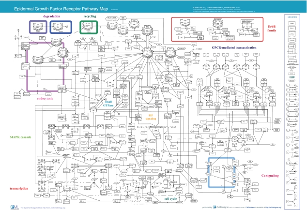
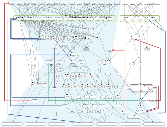
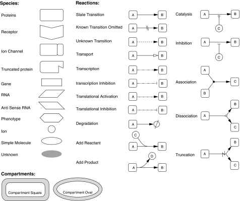
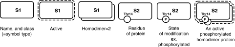
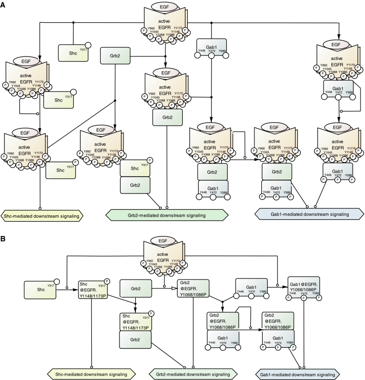
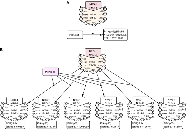
The epidermal growth factor receptor (EGFR) signaling pathway is one of the most important pathways that regulate growth, survival, proliferation, and differentiation in mammalian cells. Reflecting this importance, it is one of the best-investigated signaling systems, both experimentally and computationally, and several computational models have been developed for dynamic analysis. A map of molecular interactions of the EGFR signaling system is a valuable resource for research in this area. In this paper, we present a comprehensive pathway map of EGFR signaling and other related pathways. The map reveals that the overall architecture of the pathway is a bow-tie (or hourglass) structure with several feedback loops. The map is created using CellDesigner software that enables us to graphically represent interactions using a well-defined and consistent graphical notation, and to store it in Systems Biology Markup Language (SBML).


Figures

Figure 1
Figure 1
EGFR Pathway Map. This map was created using CellDesigner ver. 2.0 (http://www.systems-biology.org/002/). A total of 219 reactions and 322 species were included. The map can be best viewed in the PDF format. Abi, abl-interactor; ADAM, a disintegrin and metalloproteinase; ADPR, ADP-ribose; Akt, v-akt murine thymoma viral oncogene homolog; AP-1, activator protein-1; Bad, BCL2-antagonist of cell death; cADPR, cyclic ADP-ribose; CAK, cyclin-dependent kinase-activating kinase; CaM, calmodulin; CaMK, calcium/calmodulin-dependent protein kinase; c-Cbl, Casitas B-lineage lymphoma proto-oncogene; CD, cluster of differentiation; Cdc, cell division cycle; Cdk, cyclin-dependent kinase; c-Fos, v-fos FBJ murine osteosarcoma viral oncogene; Chk, c-src tyrosine kinase (Csk) homologous kinase; c-Jun, v-jun sarcoma virus 17 oncogene homolog; c-Myc, v-myc myelocytomatosis viral oncogene homolog; CREB, cAMP response element-binding protein; c-Src, v-src sarcoma viral oncogene homolog; cyt., cytosol; DAG, diacylglycerol; EGF, epidermal growth factor; EGFR, epidermal growth factor receptor; Elk, Ets-like protein; end., endosome; EP, prostaglandin E receptor; Eps, EGF receptor pathway substrate; ER, endoplasmic reticulum; ErbB, erythroblastic leukemia viral (v-erb-b) oncogene homolog; ERK, extracellular signal-regulated kinase; Gab, GRB2-associated binding protein; GPCR, G protein-coupled receptor; Grb, growth factor receptor-bound protein; HB-EGF, heparin-binding EGF-like growth factor; IP3R, inositol 1,4,5-triphosphate receptor; IP3, inositol 1,4,5-triphosphate; JNK, c-Jun N-terminal kinase; KDI, kinase domain I; LARG, leukemia-associated rho guanine nucleotide exchange factor; LIMK, LIM (Lin-11 Isl-1 Mec-3) kinase; LPA, lysophosphatidic acid; LPA1/2, lysophosphatidic acid G protein-coupled receptor 1/2; lyso., lysosome; m., messenger; MAPK, mitogen-activated protein kinase; MEKK, MAPK/ERK kinase kinase; MKK, MAP kinase kinase; MKP, MAP kinase phosphatase; MLK, mixed lineage kinase; NAD, nicotinamide adenine dinucleotide; nuc., nucleus; PAK1, p21/Cdc42/Rac1-activated kinase; PDK, 3-phosphoinositide-dependent protein kinase; PGE2, prostaglandin E2; Pi, phosphoric ion; PI3,4,5-P3, phosphatidylinositol-3,4,5-triphosphate; PI3,4-P2, phosphatidylinositol-3,4-bisphosphate; PI3K, phosphatidylinositol-3-kinase; PI4,5-P2, phosphatidylinositol-4,5-bisphosphate; PI4-P, phosphatidylinositol-4-phosphate; PI5K, phosphatidylinositol-5-kinase; PIP, phosphatidylinositol polyphosphate; PKB, protein kinase B; PKC, protein kinase C; pl.m., plasma membrane; PLC, phospholipase C; PLD, phospholipase D; PP, protein phosphatase; PTB, phosphotyrosine-binding domain; PTEN, phosphatase and tensin homolog; Pyk, proline-rich tyrosine kinase; Rab5a, RAS-associated protein RAB5a; Raf, v-raf-1 murine leukemia viral oncogene homolog; Ras, rat sarcoma viral oncogene homolog; RasGAP, Ras GTPase-activating protein; Rb, retinoblastoma; RGS, regulator of G-protein signaling; Rin, Ras interaction; RN-tre, related to the N-terminus of tre; RSK, ribosomal protein S6 kinase; RYR, ryanodine receptor; S, serine; S1P, sphingosine-1-phosphate; S1P1/2/3, sphingolipid G protein-coupled receptor 1/2/3; SERCA, sarcoplasmic/endoplasmic reticulum calcium ATPase; Shc, Src homology 2 domain containing transforming protein; SHP, Shp-2 tyrosine phosphatase; SOS, son of sevenless homolog; SPRY, Sprouty; STAT, signal transducer and activator of transcription; T, threonine; TGFα, transforming growth factor alpha; Ubc, ubiquitin-conjugating enzyme; Y, tyrosine. This image is also available as high resolution PDF (see Supplementary PDF 1) or Scalable Vector Graphic (SVG) or SBML (see Supplementary SBML 1).
Figure 2
Figure 2
The bow-tie architecture of the EGFR signaling pathway. A simplified diagram was created based on the EGFR signaling map in Figure 1. Arrows in this figure represent an informal notation of 'flow of reaction'. Various ligands bind to diverse receptor heterodimers, which then converge into a handful of molecules building a conserved core. Activities of these molecules play important roles in controlling diverse responses. Notable interactions are color-coded: red, positive feedback loop; blue, negative feedback loop; purple, inhibitory feed-forward path; green, crosstalk from GPCR cascade to EGFR cascade via calcium release. This image is also available as high resolution PDF (see Supplementary PDF 2) or Scalable Vector Graphic (SVG).
Figure 3
Figure 3
Main symbols adopted by CellDesigner ver. 2.1.1. These symbols are provided in CellDesigner ver. 2.1.1. Size and color of each module are configurable. CellDesigner also provides X-Y coordinates for each module and can distinguish between cellular compartments.
Figure 4
Figure 4
Expression of the inner structures and states. The active state of the molecule is indicated by a dashed line surrounding the molecule. State changes of a component such as phosphorylation, acetylation, ubiquitination, and allosteric changes can be represented with specific information such as target residue and position.
Figure 5
Figure 5
Ellipsis in drawing association states of proteins using an 'address'. (A) Precise association states between EGFR and adaptors. Three adaptor proteins, Shc, Grb2, and Gab1, bind to the activated EGFR via its autophosphorylated tyrosine residues. Shc binds to activated EGFR and is phosphorylated on its tyrosine 317. Grb2 binds to activated EGFR either directly or via Shc bound to activated EGFR. Gab1 also binds to activated EGFR either directly or via Grb2 bound to activated EGFR, and is phosphorylated on its tyrosine 446, 472, and 589. (B) The same signaling pathway as in panel A using an 'address' such as 'Grb2@EGFR.Y1068/1086P', thereby achieving a presentation of the pathway details. EGF, epidermal growth factor; EGFR, epidermal growth factor receptor; Gab, GRB2-associated binding protein; Grb, growth factor receptor-bound protein; Shc, Src homology 2 domain containing transforming protein.
Figure 6
Figure 6
Combinations of multiple states of complexes. (A) Omitted notation in the EGFR Pathway Map. The 85 kDa regulatory subunit of phosphatidylinositol 3-kinase (PI3K (p85)) binds to active ErbB3 receptor via its phosphorylated tyrosine residues: Tyr1035, Tyr1178, Tyr1203/05, Tyr1241, Tyr1257, and Tyr1270. (B) Detailed portrayal of panel A for distinguishing the complexes according to differences of phosphotyrosine residues. ErbB3, erythroblastic leukemia viral (v-erb-b) oncogene homolog 3; PI3K (p85), 85 kDa regulatory subunit of phosphatidylinositol 3-kinase.

Comment in

References

    1. Agazie YM, Hayman MJ (2003) Molecular mechanism for a role of SHP2 in epidermal growth factor receptor signaling. Mol Cell Biol 23: 7875–7886 - PMC - PubMed
    1. Carpenter G (2000) EGF receptor transactivation mediated by the proteolytic production of EGF-like agonists. Sci STKE 2000: PE1 - PubMed
    1. Chen X, Levkowitz G, Tzahar E, Karunagaran D, Lavi S, Ben-Baruch N, Leitner O, Ratzkin BJ, Bacus SS, Yarden Y (1996) An immunological approach reveals biological differences between the two NDF/heregulin receptors, ErbB-3 and ErbB-4. J Biol Chem 271: 7620–7629 - PubMed
    1. Christoforidis S, Miaczynska M, Ashman K, Wilm M, Zhao L, Yip SC, Waterfield MD, Backer JM, Zerial M (1999) Phosphatidylinositol-3-OH kinases are Rab5 effectors. Nat Cell Biol 1: 249–252 - PubMed
    1. Cook DL, Farley JF, Tapscott SJ (2001) A basis for a visual language for describing, archiving and analyzing functional models of complex biological systems. Genome Biol 2, RESEARCH0012 - PMC - PubMed

[References for EGFR Pathway Map] Epidermal growth factor (EGFR)

    1. Agazie YM, Hayman MJ (2003) Molecular mechanism for a role of SHP2 in epidermal growth factor receptor signaling. Mol Cell Biol 23: 7875–7886 - PMC - PubMed
    1. Avraham H, Park SY, Schinkmann K, Avraham S (2000) RAFTK/Pyk2-mediated cellular signalling. Cell Signal 12: 123–133 - PubMed
    1. Batzer AG, Rotin D, Urena JM, Skolnik EY, Schlessinger J (1994) Hierarchy of binding sites for Grb2 and Shc on the epidermal growth factor receptor. Mol Cell Biol 14: 5192–5201 - PMC - PubMed
    1. Biscardi JS, Maa MC, Tice DA, Cox ME, Leu TH, Parsons SJ (1999) c-Src-mediated phosphorylation of the epidermal growth factor receptor on Tyr845 and Tyr1101 is associated with modulation of receptor function. J Biol Chem 274: 8335–8343 - PubMed
    1. Bishayee A, Beguinot L, Bishayee S (1999) Phosphorylation of tyrosine 992, 1068, and 1086 is required for conformational change of the human epidermal growth factor receptor C-terminal tail. Mol Biol Cell 10: 525–536 - PMC - PubMed

EGFR endocytosis followed by degradation or recycling

    1. Bao J, Alroy I, Waterman H, Schejter ED, Brodie C, Gruenberg J, Yarden Y (2000) Threonine phosphorylation diverts internalized epidermal growth factor receptors from a degradative pathway to the recycling endosome. J Biol Chem 275: 26178–26186 - PubMed
    1. Barbieri MA, Roberts RL, Gumusboga A, Highfield H, Alvarez-Dominguez C, Wells A, Stahl PD (2000) Epidermal growth factor and membrane trafficking. EGF receptor activation of endocytosis requires Rab5a. J Cell Biol 151: 539–550 - PMC - PubMed
    1. Han L, Wong D, Dhaka A, Afar D, White M, Xie W, Herschman H, Witte O, Colicelli J (1997) Protein binding and signaling properties of RIN1 suggest a unique effector function. Proc Natl Acad Sci USA 94: 4954–4959 - PMC - PubMed
    1. Jiang X, Sorkin A (2002) Coordinated traffic of Grb2 and Ras during epidermal growth factor receptor endocytosis visualized in living cells. Mol Biol Cell 13: 1522–1535 - PMC - PubMed
    1. Kassenbrock CK, Hunter S, Garl P, Johnson GL, Anderson SM (2002) Inhibition of Src family kinases blocks epidermal growth factor (EGF)-induced activation of Akt, phosphorylation of c-Cbl, and ubiquitination of the EGF receptor. J Biol Chem 277: 24967–24975 - PubMed

Small GTPase

    1. Ahmed S, Lee J, Kozma R, Best A, Monfries C, Lim L (1993) A novel functional target for tumor-promoting phorbol esters and lysophosphatidic acid. The p21rac-GTPase activating protein n-chimaerin. J Biol Chem 268: 10709–10712 - PubMed
    1. Burbelo PD, Drechsel D, Hall A (1995) A conserved binding motif defines numerous candidate target proteins for both Cdc42 and Rac GTPases. J Biol Chem 270: 29071–29074 - PubMed
    1. Caloca MJ, Wang H, Delemos A, Wang S, Kazanietz MG (2001) Phorbol esters and related analogs regulate the subcellular localization of beta 2-chimaerin, a non-protein kinase C phorbol ester receptor. J Biol Chem 276: 18303–18312 - PubMed
    1. Caloca MJ, Wang H, Kazanietz MG (2003) Characterization of the Rac-GAP (Rac-GTPase-activating protein) activity of beta2-chimaerin, a 'non-protein kinase C' phorbol ester receptor. Biochem J 375: 313–321 - PMC - PubMed
    1. Cox AD, Der CJ (2003) The dark side of Ras: regulation of apoptosis. Oncogene 22: 8999–9006 - PubMed

Phosphatidylinositol phosphate (PIP) signaling

    1. Alessi DR, Deak M, Casamayor A, Caudwell FB, Morrice N, Norman DG, Gaffney P, Reese CB, MacDougall CN, Harbison D, Ashworth A, Bownes M (1997) 3-Phosphoinositide-dependent protein kinase-1 (PDK1): structural and functional homology with the Drosophila DSTPK61 kinase. Curr Biol 7: 776–789 - PubMed
    1. Belham C, Wu S, Avruch J (1999) Intracellular signalling: PDK1–a kinase at the hub of things. Curr Biol 9: R93–R96 - PubMed
    1. Carpenter G, Ji Q (1999) Phospholipase C-gamma as a signal-transducing element. Exp Cell Res 253: 15–24 - PubMed
    1. Casamayor A, Morrice NA, Alessi DR (1999) Phosphorylation of Ser-241 is essential for the activity of 3-phosphoinositide-dependent protein kinase-1: identification of five sites of phosphorylation in vivo . Biochem J 342: 287–292 - PMC - PubMed
    1. Datta SR, Dudek H, Tao X, Masters S, Fu H, Gotoh Y, Greenberg ME (1997) Akt phosphorylation of BAD couples survival signals to the cell-intrinsic death machinery. Cell 91: 231–241 - PubMed

Mitogen-activated protein kinase (MAPK) cascade

    1. Carey KD, Watson RT, Pessin JE, Stork PJ (2003) The requirement of specific membrane domains for Raf-1 phosphorylation and activation. J Biol Chem 278: 3185–3196 - PubMed
    1. Chi H, Sarkisian MR, Rakic P, Flavell RA (2005) Loss of mitogen-activated protein kinase kinase kinase 4 (MEKK4) results in enhanced apoptosis and defective neural tube development. Proc Natl Acad Sci USA 102: 3846–3851 - PMC - PubMed
    1. Chrestensen CA, Sturgill TW (2002) Characterization of the p90 ribosomal S6 kinase 2 carboxyl-terminal domain as a protein kinase. J Biol Chem 277: 27733–27741 - PubMed
    1. Cleghon V, Morrison DK (1994) Raf-1 interacts with Fyn and Src in a non-phosphotyrosine-dependent manner. J Biol Chem 269: 17749–17755 - PubMed
    1. Crews CM, Alessandrini A, Erikson RL (1992) The primary structure of MEK, a protein kinase that phosphorylates the ERK gene product. Science 258: 478–480 - PubMed

Transcription

    1. Alvarez E, Northwood IC, Gonzalez FA, Latour DA, Seth A, Abate C, Curran T, Davis RJ (1991) Pro-Leu-Ser/Thr-Pro is a consensus primary sequence for substrate protein phosphorylation. Characterization of the phosphorylation of c-myc and c-jun proteins by an epidermal growth factor receptor threonine 669 protein kinase. J Biol Chem 266: 15277–15285 - PubMed
    1. Aplin AE, Stewart SA, Assoian RK, Juliano RL (2001) Integrin-mediated adhesion regulates ERK nuclear translocation and phosphorylation of Elk-1. J Cell Biol 153: 273–282 - PMC - PubMed
    1. Cavigelli M, Dolfi F, Claret FX, Karin M (1995) Induction of c-fos expression through JNK-mediated TCF/Elk-1 phosphorylation. EMBO J 14: 5957–5964 - PMC - PubMed
    1. Coronella-Wood J, Terrand J, Sun H, Chen QM (2004) c-Fos phosphorylation induced by H2O2 prevents proteasomal degradation of c-Fos in cardiomyocytes. J Biol Chem 279: 33567–33574 - PubMed
    1. De Cesare D, Jacquot S, Hanauer A, Sassone-Corsi P (1998) Rsk-2 activity is necessary for epidermal growth factor-induced phosphorylation of CREB protein and transcription of c-fos gene. Proc Natl Acad Sci USA 95: 12202–12207 - PMC - PubMed

Cell cycle

    1. Alkarain A, Jordan R, Slingerland J (2004) p27 deregulation in breast cancer: prognostic significance and implications for therapy. J Mammary Gland Biol Neoplasia 9: 67–80 - PubMed
    1. Bakiri L, Lallemand D, Bossy-Wetzel E, Yaniv M (2000) Cell cycle-dependent variations in c-Jun and JunB phosphorylation: a role in the control of cyclin D1 expression. EMBO J 19: 2056–2068 - PMC - PubMed
    1. Bartkova J, Rajpert-De Meyts E, Skakkebaek NE, Lukas J, Bartek J (2003) Deregulation of the G1/S-phase control in human testicular germ cell tumours. Apmis 111: 252–265, discussion 265–256 - PubMed
    1. Bates S, Bonetta L, MacAllan D, Parry D, Holder A, Dickson C, Peters G (1994) CDK6 (PLSTIRE) and CDK4 (PSK-J3) are a distinct subset of the cyclin-dependent kinases that associate with cyclin D1. Oncogene 9: 71–79 - PubMed
    1. Botz J, Zerfass-Thome K, Spitkovsky D, Delius H, Vogt B, Eilers M, Hatzigeorgiou A, Jansen-Durr P (1996) Cell cycle regulation of the murine cyclin E gene depends on an E2F binding site in the promoter. Mol Cell Biol 16: 3401–3409 - PMC - PubMed

G protein-coupled receptor (GPCR)-mediated EGFR transactivation

    1. Beebe SJ (1994) The cAMP-dependent protein kinases and cAMP signal transduction. Semin Cancer Biol 5: 285–294 - PubMed
    1. Benzing T, Yaffe MB, Arnould T, Sellin L, Schermer B, Schilling B, Schreiber R, Kunzelmann K, Leparc GG, Kim E, Walz G (2000) 14-3-3 interacts with regulator of G protein signaling proteins and modulates their activity. J Biol Chem 275: 28167–28172 - PubMed
    1. Colley WC, Sung TC, Roll R, Jenco J, Hammond SM, Altshuller Y, Bar-Sagi D, Morris AJ, Frohman MA (1997) Phospholipase D2, a distinct phospholipase D isoform with novel regulatory properties that provokes cytoskeletal reorganization. Curr Biol 7: 191–201 - PubMed
    1. Dzimiri N (2002) Receptor crosstalk. Implications for cardiovascular function, disease and therapy. Eur J Biochem 269: 4713–4730 - PubMed
    1. Exton JH (2002) Regulation of phospholipase D. FEBS Lett 531: 58–61 - PubMed

Ca2+ signaling

    1. Banno Y, Asano T, Nozawa Y (1994) Proteolytic modification of membrane-associated phospholipase C-beta by mu-calpain enhances its activation by G-protein beta gamma subunits in human platelets. FEBS Lett 340: 185–188 - PubMed
    1. Bruce JI, Shuttleworth TJ, Giovannucci DR, Yule DI (2002) Phosphorylation of inositol 1,4,5-trisphosphate receptors in parotid acinar cells. A mechanism for the synergistic effects of cAMP on Ca2+ signaling. J Biol Chem 277: 1340–1348 - PubMed
    1. Danila CI, Hamilton SL (2004) Phosphorylation of ryanodine receptors. Biol Res 37: 521–525 - PubMed
    1. DeSouza N, Reiken S, Ondrias K, Yang YM, Matkovich S, Marks AR (2002) Protein kinase A and two phosphatases are components of the inositol 1,4,5-trisphosphate receptor macromolecular signaling complex. J Biol Chem 277: 39397–39400 - PubMed
    1. Ishida A, Shigeri Y, Taniguchi T, Kameshita I (2003) Protein phosphatases that regulate multifunctional Ca2+/calmodulin-dependent protein kinases: from biochemistry to pharmacology. Pharmacol Ther 100: 291–305 - PubMed

ErbB family

    1. Cohen BD, Green JM, Foy L, Fell HP (1996) HER4-mediated biological and biochemical properties in NIH 3T3 cells. Evidence for HER1-HER4 heterodimers. J Biol Chem 271: 4813–4818 - PubMed
    1. Elenius K, Choi CJ, Paul S, Santiestevan E, Nishi E, Klagsbrun M (1999) Characterization of a naturally occurring ErbB4 isoform that does not bind or activate phosphatidyl inositol 3-kinase. Oncogene 18: 2607–2615 - PubMed
    1. Fiddes RJ, Campbell DH, Janes PW, Sivertsen SP, Sasaki H, Wallasch C, Daly RJ (1998) Analysis of Grb7 recruitment by heregulin-activated erbB receptors reveals a novel target selectivity for erbB3. J Biol Chem 273: 7717–7724 - PubMed
    1. Hazan R, Margolis B, Dombalagian M, Ullrich A, Zilberstein A, Schlessinger J (1990) Identification of autophosphorylation sites of HER2/neu. Cell Growth Differ 1: 3–7 - PubMed
    1. Prigent SA, Gullick WJ (1994) Identification of c-erbB-3 binding sites for phosphatidylinositol 3′-kinase and SHC using an EGF receptor/c-erbB-3 chimera. EMBO J 13: 2831–2841 - PMC - PubMed

Publication types

Substances