

The expression of p63 is associated with the differential stage in nasopharyngeal carcinoma and EBV infection
- PMID: 16729897
- PMCID: PMC1525203
- DOI: 10.1186/1479-5876-4-23
The expression of p63 is associated with the differential stage in nasopharyngeal carcinoma and EBV infection
Abstract
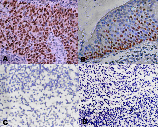
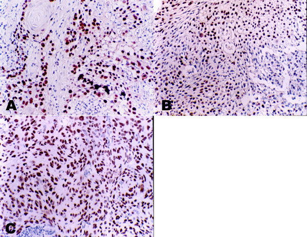
Background: Nasopharyngeal carcinoma (NPC) is common among Southern Chinese and the main histology is the undifferentiated carcinoma associated with Epstein-Barr virus (EBV) infection. p63 is a recently proved member of the p53 family based on the structural similarity to p53, but its function in NPC is still unknown. This study was aimed to investigate the association between p63 and NPC.
Results: p63 was expressed in 100% (202/202) of nasopharyngeal carcinoma (NPC) tissues but not in 29 nasopharynx inflammation and 17 non-cancerous nasopharyngeal epidermises on a tissue microarray by immunohistostaining. Further investigation suggested that the p63 expression was associated with the differential stage of NPC: p63 strong staining in Keratinizing squamous cell carcinoma, differentiated non-keratinizing NPC and undifferentiated non-keratinizing NPC presented the percentage of 5/8 (62.5%), 43/48 (92.5%) and 50/50 (100%), respectively. A significant difference (p = 0.001) existed between the keratinizing and non-keratinizing groups. No pathogenic mutations were detected in p63 gene in 12 primary NPC tissues and matched peripheral blood lymphocytes (PBL). Half-life measurement study revealed distinct stability of p63 protein in the different cell lines, especially between the carcinoma cell lines with EBV infection and the non-cancerous cell lines. The results of immunoprecipitation suggested a direct interaction between Epstein-Barr virus nuclear antigen 5 (EBNA-5) and p63 protein in NPC, and this binding would increase the stability of p63.
Conclusion: Our data suggested p63 might be used as an adjunct diagnostic marker of NPC and contributed a new way to understand the contribution of the EBV in the pathogenesis of NPC.
Figures


Similar articles
-
[Expression of CD25+ lymphocytes in nasopharyngeal carcinoma and its association with EBV infection].Nan Fang Yi Ke Da Xue Xue Bao. 2006 Jan;26(1):94-7. Nan Fang Yi Ke Da Xue Xue Bao. 2006. PMID: 16495186 Chinese.
-
Hypermethylation of epithelial-cadherin gene promoter is associated with Epstein-Barr virus in nasopharyngeal carcinoma.Cancer Detect Prev. 2008;32(2):127-34. doi: 10.1016/j.cdp.2008.05.005. Epub 2008 Jul 15. Cancer Detect Prev. 2008. PMID: 18632221
-
Comparison of Epstein-Barr virus genotypes and clinicohistopathological features of nasopharyngeal carcinoma between Guilin, China and Fukuoka, Japan.Oncol Rep. 2008 Jun;19(6):1413-20. Oncol Rep. 2008. PMID: 18497945
-
Mechanisms of cell immortalization mediated by EB viral activation of telomerase in nasopharyngeal carcinoma.Cell Res. 2006 Oct;16(10):809-17. doi: 10.1038/sj.cr.7310098. Cell Res. 2006. PMID: 17016469 Review.
-
Nasopharyngeal carcinoma among the Bidayuh of Sarawak, Malaysia: History and risk factors.Oncol Lett. 2021 Jul;22(1):514. doi: 10.3892/ol.2021.12775. Epub 2021 May 6. Oncol Lett. 2021. PMID: 33986874 Free PMC article. Review.
Cited by
-
The interplay of host genetic factors and Epstein-Barr virus in the development of nasopharyngeal carcinoma.Chin J Cancer. 2014 Nov;33(11):556-68. doi: 10.5732/cjc.014.10170. Chin J Cancer. 2014. PMID: 25367335 Free PMC article. Review.
-
The interplay between Epstein-Bar virus (EBV) with the p53 and its homologs during EBV associated malignancies.Heliyon. 2019 Nov 14;5(11):e02624. doi: 10.1016/j.heliyon.2019.e02624. eCollection 2019 Nov. Heliyon. 2019. PMID: 31840114 Free PMC article. Review.
-
Apogossypolone targets mitochondria and light enhances its anticancer activity by stimulating generation of singlet oxygen and reactive oxygen species.Chin J Cancer. 2011 Jan;30(1):41-53. doi: 10.5732/cjc.010.10295. Chin J Cancer. 2011. PMID: 21192843 Free PMC article.
-
circTP63-N suppresses the proliferation and metastasis of nasopharyngeal carcinoma via engaging with HSP90AB1 to modulate the YAP1/Hippo signaling pathway.Sci China Life Sci. 2025 Mar;68(3):689-705. doi: 10.1007/s11427-023-2737-2. Epub 2024 Dec 27. Sci China Life Sci. 2025. PMID: 39754006
-
Molecular classification and molecular targeted therapy of cancer.Front Med. 2013 Jun;7(2):147-9. doi: 10.1007/s11684-013-0274-2. Front Med. 2013. PMID: 23686608 No abstract available.
References
-
- Hildesheim A, West S, DeVeyra E, De Guzman MF, Jurado A, Jones C, Imai J, Hinuma Y. Herbal medicine use, Epstein-Barr virus, and risk of nasopharyngeal carcinoma. Cancer Res. 1992;52:3048–3051. - PubMed
-
- Raab-Traub N. Epstein-Barr virus and nasopharyngeal carcinoma. Semin Cancer Biol. 1992;3:297–307. - PubMed
-
- Xiong W, Zeng ZY, Xia JH, Xia K, Shen SR, Li XL, Hu DX, Tan C, Xiang JJ, Zhou J, Deng H, Fan SQ, Li WF, Wang R, Zhou M, Zhu SG, Lu HB, Qian J, Zhang BC, Wang JR, Ma J, Xiao BY, Huang H, Zhang QH, Zhou YH, Luo XM, Zhou HD, Yang YX, Dai HP, Feng GY, Pan Q, Wu LQ, He L, Li GY. A susceptibility locus at chromosome 3p21 linked to familial nasopharyngeal carcinoma. Cancer Res. 2004;64:1972–1974. doi: 10.1158/0008-5472.CAN-03-3253. - DOI - PubMed
LinkOut - more resources
Full Text Sources
Research Materials
Miscellaneous
