Biomechanical signals exert sustained attenuation of proinflammatory gene induction in articular chondrocytes
- PMID: 16731008
- PMCID: PMC4950917
- DOI: 10.1016/j.joca.2006.03.016
Biomechanical signals exert sustained attenuation of proinflammatory gene induction in articular chondrocytes
Abstract
Objectives: Physical therapies are commonly used for limiting joint inflammation. To gain insight into their mechanisms of actions for optimal usage, we examined persistence of mechanical signals generated by cyclic tensile strain (CTS) in chondrocytes, in vitro. We hypothesized that mechanical signals induce anti-inflammatory and anabolic responses that are sustained over extended periods.
Methods: Articular chondrocytes obtained from rats were subjected to CTS for various time intervals followed by a period of rest, in the presence of interleukin-1beta (IL-1beta). The induction for cyclooxygenase (COX-2), inducible nitric oxide synthase (iNOS), matrix metalloproteinase (MMP)-9, MMP-13 and aggrecan was analyzed by real-time polymerase chain reaction (PCR), Western blot analysis and immunofluorescence.
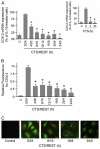
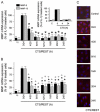
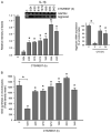
Results: Exposure of chondrocytes to constant CTS (3% CTS at 0.25 Hz) for 4-24 h blocked more than 90% (P<0.05) of the IL-1beta-induced transcriptional activation of proinflammatory genes, like iNOS, COX-2, MMP-9 and MMP-13, and abrogated inhibition of aggrecan synthesis. CTS exposure for 4, 8, 12, 16, or 20 h followed by a rest for 20, 16, 12, 8 or 4h, respectively, revealed that 8h of CTS optimally blocked (P<0.05) IL-1beta-induced proinflammatory gene induction for ensuing 16 h. However, CTS for 8h was not sufficient to inhibit iNOS expression for ensuing 28 or 40 h.
Conclusions: Data suggest that constant application of CTS blocks IL-1beta-induced proinflammatory genes at transcriptional level. The signals generated by CTS are sustained after its removal, and their persistence depends upon the length of CTS exposure. Furthermore, the sustained effects of mechanical signals are also reflected in their ability to induce aggrecan synthesis. These findings, once extrapolated to human chondrocytes, may provide insight in obtaining optimal sustained effects of physical therapies in the management of arthritic joints.
Figures


Similar articles
-
Cyclic tensile strain suppresses catabolic effects of interleukin-1beta in fibrochondrocytes from the temporomandibular joint.Arthritis Rheum. 2001 Mar;44(3):608-17. doi: 10.1002/1529-0131(200103)44:3<608::AID-ANR109>3.0.CO;2-2. Arthritis Rheum. 2001. PMID: 11263775 Free PMC article.
-
Cyclic tensile strain acts as an antagonist of IL-1 beta actions in chondrocytes.J Immunol. 2000 Jul 1;165(1):453-60. doi: 10.4049/jimmunol.165.1.453. J Immunol. 2000. PMID: 10861084 Free PMC article.
-
Sclareol exerts anti-osteoarthritic activities in interleukin-1β-induced rabbit chondrocytes and a rabbit osteoarthritis model.Int J Clin Exp Pathol. 2015 Mar 1;8(3):2365-74. eCollection 2015. Int J Clin Exp Pathol. 2015. PMID: 26045743 Free PMC article.
-
Regulation of biomechanical signals by NF-kappaB transcription factors in chondrocytes.Biorheology. 2008;45(3-4):245-56. Biorheology. 2008. PMID: 18836228 Free PMC article. Review.
-
Regulation of chondrocytic gene expression by biomechanical signals.Crit Rev Eukaryot Gene Expr. 2008;18(2):139-50. doi: 10.1615/critreveukargeneexpr.v18.i2.30. Crit Rev Eukaryot Gene Expr. 2008. PMID: 18304028 Free PMC article. Review.
Cited by
-
GREM1, FRZB and DKK1 mRNA levels correlate with osteoarthritis and are regulated by osteoarthritis-associated factors.Arthritis Res Ther. 2013 Sep 19;15(5):R126. doi: 10.1186/ar4306. Arthritis Res Ther. 2013. PMID: 24286177 Free PMC article.
-
Role of melatonin combined with exercise as a switch-like regulator for circadian behavior in advanced osteoarthritic knee.Oncotarget. 2017 Jul 16;8(57):97633-97647. doi: 10.18632/oncotarget.19276. eCollection 2017 Nov 14. Oncotarget. 2017. PMID: 29228639 Free PMC article.
-
The basic science of continuous passive motion in promoting knee health: a systematic review of studies in a rabbit model.Arthroscopy. 2013 Oct;29(10):1722-31. doi: 10.1016/j.arthro.2013.05.028. Epub 2013 Jul 26. Arthroscopy. 2013. PMID: 23890952 Free PMC article.
-
Mechanisms Underlying Anti-Inflammatory and Anti-Cancer Properties of Stretching-A Review.Int J Mol Sci. 2022 Sep 4;23(17):10127. doi: 10.3390/ijms231710127. Int J Mol Sci. 2022. PMID: 36077525 Free PMC article. Review.
-
Mechanosignaling in bone health, trauma and inflammation.Antioxid Redox Signal. 2014 Feb 20;20(6):970-85. doi: 10.1089/ars.2013.5467. Epub 2013 Aug 12. Antioxid Redox Signal. 2014. PMID: 23815527 Free PMC article. Review.
References
-
- Buckwalter JA, Mankin HJ, Grodzinsky AJ. Articular cartilage and osteoarthritis. Instr Course Lect. 2005;54:465–80. - PubMed
-
- Kurz B, Lemke AK, Fay J, Pufe T, Grodzinsky AJ, Schunke M. Pathomechanisms of cartilage destruction by mechanical injury. Ann Anat. 2005;187:473–85. - PubMed
-
- Milne S, Brosseau L, Robinson V, Noel MJ, Davis J, Drouin H, et al. Continuous passive motion following total knee arthroplasty. Cochrane Database Syst Rev. 2003;2:CD004260. - PubMed
-
- Griffin TM, Guilak F. The role of mechanical loading in the onset and progression of osteoarthritis. Exerc Sport Sci Rev. 2005;33:195–200. - PubMed
-
- Das UN. Anti-inflammatory nature of exercise. Nutrition. 2004;20:323–6. - PubMed
Publication types
MeSH terms
Substances
Grants and funding
LinkOut - more resources
Full Text Sources
Research Materials
Miscellaneous

