. 2006 Sep 1;91(5):1960-9.
doi: 10.1529/biophysj.106.082420. Epub 2006 May 26.

Atomic force microscopy study of the specific adhesion between a colloid particle and a living melanoma cell: Effect of the charge and the hydrophobicity of the particle surface

Affiliations

Atomic force microscopy study of the specific adhesion between a colloid particle and a living melanoma cell: Effect of the charge and the hydrophobicity of the particle surface

Cathy E McNamee et al. Biophys J. .

Abstract

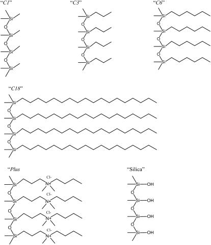
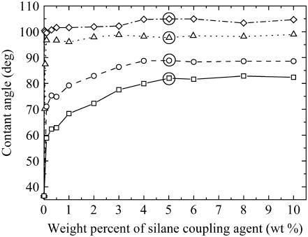
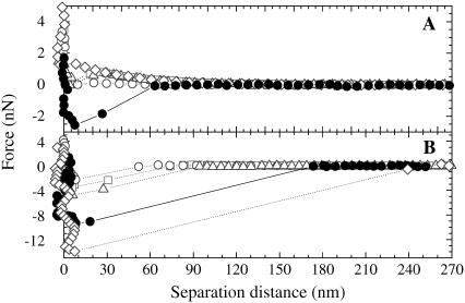
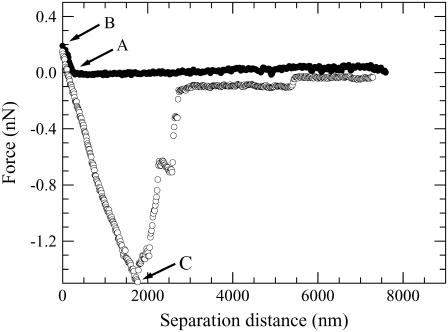
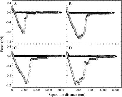
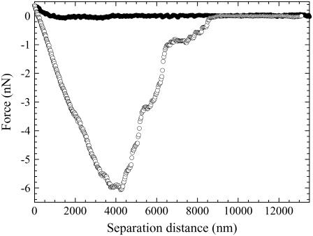
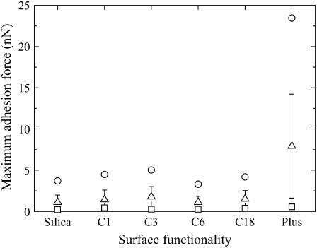
We investigated the effect of the charge and the hydrophobicity of drug delivery system (DDS) carriers on their specificity to living malignant melanoma B16F10 cells with the atomic force microscope. To model various nanoparticle DDS carriers, we used silica particles that were modified with silane coupling agents. We then measured the compression and decompression forces between the modified colloid probes and the living B16F10 cell in a physiological buffer as a function of their separation distances. The maximum adhesive force on decompression was related to the strength of the specificity of the DDS to the malignant cell. A comparison of the average maximum adhesive force of each functionality group surprisingly showed that negatively charged surfaces and hydrophobic modified surfaces all had similar low values. Additionally, we saw the unexpected result that there was no observable dependence on the degree of hydrophobicity of the probe surface to a B16F10 cell. Only the positively charged particle gave a strong adhesive force with the B16F10 cell. This indicated that DDS carriers with positive charges appeared to have the highest affinity for malignant melanoma cells and that the use of hydrophobic materials unexpectedly did not improve their affinity.


Figures

FIGURE 1
FIGURE 1
A schematic picture depicting the surface of the modified particles. C1, C3, C6, C18, and Plus refer to silica surfaces modified by trimethoxy(methyl)silane, trimethoxy-n-propylsilane, n-hexyltrimethoxysilane, octadecyltrimethoxysilane, and N-trimethylsilylpropyl-N,N,N-trimethylammonium chloride, respectively. Silica shows the surface groups on a silica particle.
FIGURE 2
FIGURE 2
The change in the contact angle of a glass substrate as a function of the concentration of silane coupling agents when functionalized in the method given in the experimental section. Here the different silane coupling agents are depicted as C1 (□ with solid line); C3 (○ with dashed line); C6 (Δ with dotted line); and C18 (◊ with dashed-dotted line). The maximum adsorption of the silane coupling agents commences at the horizontal plateau in the contact angle versus concentration data. A solid circle is drawn around the concentrations used to functionalize the silica particles.
FIGURE 3
FIGURE 3
The forces between the functionalized silica particles and a freshly cleaved, mica plate in water of pH 5.6. Here, we used C1- (□ with dotted line), C3- (○ with dotted line), C6- (Δ with dotted line), C18- (◊ with dotted line), and Plus-modified (• with solid line) silica particles. Panels A and B show the compression and decompression forces, respectively.
FIGURE 4
FIGURE 4
Light microscope image of living B16F10 cells adsorbed at bare and functionalized glass substrates. Panels A, B, C, D, E, and F show the glass (model for silica), Plus, C1, C3, C6, and C18 surfaces, respectively.
FIGURE 5
FIGURE 5
Light microscope image of dead B16F10 cells adsorbed at bare and functionalized glass substrates. Panels A, B, C, D, E, and F show the glass (model for silica), Plus, C1, C3, C6, and C18 surfaces, respectively.
FIGURE 6
FIGURE 6
An example of a force curve measured between a bare silica particle (negative charge) and a B16F10 cell in the L-15 solution. The decompression force curve was measured after the particle resided at the cell surface for 10 min. Points A, B, and C depict the surface of the cell, the position relative to the cell surface where the particle resided for 10 min, and the maximum adhesion force, respectively.
FIGURE 7
FIGURE 7
Examples of the forces obtained between (A) C1-, (B) C3-, (C) C6-, and (D) C18-modified colloid probes and a B16F10 cell in the L-15 solution. The decompression force curve was measured after the particle resided at the cell surface for 10 min.
FIGURE 8
FIGURE 8
An example of a force curve measured between a positively charged particle (a silica particle modified with Plus) and the B16F10 cell in the L-15 solution. The decompression force curve was measured after the particle resided at the cell surface for 10 min.
FIGURE 9
FIGURE 9
The effect of the different functionality groups on their adhesive strength to the B16F10 cell. Here, the maximum (○), minimum (▵), and average values obtained for the maximum adhesive force (□) and its standard deviation (solid line) are compared for each functionality group (x axis).

References

    1. Ferrari, M. 2005. Cancer nanotechnology: opportunities and challenges. Nat. Rev. Cancer. 5:161–171. - PubMed
    1. Muller-Goymann, C. C. 2004. Physiochemical characterization of colloidal drug delivery systems such as reverse micelles, vesicles, liquid crystals and nanoparticles for topical administration. Eur. J. Pharm. Biopharm. 58:343–356. - PubMed
    1. Alberts, A., A. Johnson, J. Lewis, M. Raff, K. Roberts, and P. Walter. 2002. Molecular Biology of the Cell. Garland Science, New York. 376.
    1. Alberts, A., A. Johnson, J. Lewis, M. Raff, K. Roberts, and P. Walter. 2002. Molecular Biology of the Cell. Garland Science, New York. 1314.
    1. Campisi, J. 2000. Cancer, aging and cellular sequence. In Vivo. 14:183–188. - PubMed

Publication types

LinkOut - more resources