. 2006 Jul;18(7):1693-703.
doi: 10.1105/tpc.105.039859. Epub 2006 May 26.

Plastoglobules are lipoprotein subcompartments of the chloroplast that are permanently coupled to thylakoid membranes and contain biosynthetic enzymes

Affiliations

Plastoglobules are lipoprotein subcompartments of the chloroplast that are permanently coupled to thylakoid membranes and contain biosynthetic enzymes

Jotham R Austin 2nd et al. Plant Cell. 2006 Jul.

Abstract

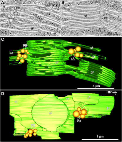
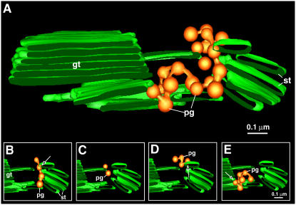
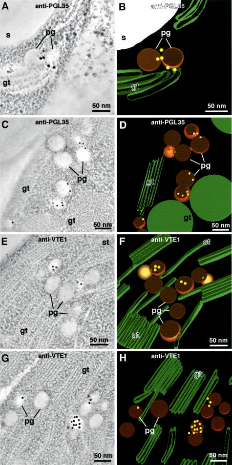
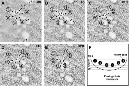
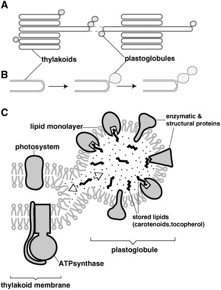
Plastoglobules are lipoprotein particles inside chloroplasts. Their numbers have been shown to increase during the upregulation of plastid lipid metabolism in response to oxidative stress and during senescence. In this study, we used state-of-the-art high-pressure freezing/freeze-substitution methods combined with electron tomography as well as freeze-etch electron microscopy to characterize the structure and spatial relationship of plastoglobules to thylakoid membranes in developing, mature, and senescing chloroplasts. We demonstrate that plastoglobules are attached to thylakoids through a half-lipid bilayer that surrounds the globule contents and is continuous with the stroma-side leaflet of the thylakoid membrane. During oxidative stress and senescence, plastoglobules form linkage groups that are attached to each other and remain continuous with the thylakoid membrane by extensions of the half-lipid bilayer. Using three-dimensional tomography combined with immunolabeling techniques, we show that the plastoglobules contain the enzyme tocopherol cyclase (VTE1) and that this enzyme extends across the surface monolayer into the interior of the plastoglobules. These findings demonstrate that plastoglobules function as both lipid biosynthesis and storage subcompartments of thylakoid membranes. The permanent structural coupling between plastoglobules and thylakoid membranes suggests that the lipid molecules contained in the plastoglobule cores (carotenoids, plastoquinone, and tocopherol [vitamin E]) are in a dynamic equilibrium with those located in the thylakoid membranes.


Figures

Figure 1.
Figure 1.
Plastoglobule Location in Relation to the Thylakoid Membranes. (A) and (B) Two composite tomographic slice images (five superimposed serial 2.2-nm slices) of grana thylakoids (gt), stroma thylakoids (st), and plastoglobules (pg) in an intact, isolated spinach chloroplast. (C) and (D) Tomographic model of the grana stack shown in (A) and (B) as seen in a side view (C) and a top-down view (D) ([C] rotated 90°). Note that in (D), the top-most stoma thylakoid has been removed to provide a clearer view of the clusters of plastoglobules that are associated with areas of high curvature of the stroma and grana thylakoids.
Figure 2.
Figure 2.
Electron Tomographic Imaging of Plastoglobule–Thylakoid Membrane Connecting Sites. (A) to (C) Serial 2.2-nm tomographic slice images (every fourth slice shown) through a plastoglobule (pg) blistering from the margin of a thylakoid membrane from a greening Arabidopsis chloroplast. The arrowheads point to the sites where the outer leaflet of the thylakoid membrane has separated from the inner leaflet to form the half bilayer surrounding the plastoglobule. Note that the inner leaflet of the thylakoid membrane can also be seen in (B). gt, grana thylakoid; st, stroma thylakoid. (D) Tomographic model of the plastoglobule shown in (A) to (C). (E) A face-on view of a plastoglobule–thylakoid membrane connection. (F) to (H) Serial 2.2-nm rotated (x = 20°, y = 21.5°, z = 6.5°) tomographic slice images of (E) through a plastoglobule–thylakoid connection. Note that the arrowheads in (G) point to the neck-like plastoglobule–thylakoid membrane continuous membrane connection.
Figure 3.
Figure 3.
Freeze-Fracture Electron Micrographs of Plastoglobule–Thylakoid and Plastoglobule–Plastoglobule Connections. (A) and (B) Two examples of plastoglobule–thylakoid interactions. In both images, the half-lipid bilayer that surrounds the lipid core has been partly torn away to expose the lipidic contents of the plastoglobule (pg). The edge of the remaining cross-fractured half bilayer is seen as a ridge-like structure that can be followed through the narrow neck region to the stroma leaflet of the thylakoid membrane (arrowheads). The extension of the core material in the neck region up to the middle of the thylakoid membrane is most evident in (A) (cf. also with Figure 2G). (C) The arrowhead points to a cross-fractured neck region exposed by the breaking away of the plastoglobule. The neck region, as seen from the inside of the plastoglobule, exhibits a funnel-shaped geometry. (D) Longitudinal fracture through two plastoglobules connected through a half-lipid bilayer tube.
Figure 4.
Figure 4.
Interconnected Plastoglobules. Two composite tomographic slice images ([A] and [C]) (five superimposed serial 2.2-nm slices) of groups of interconnected plastoglobules held together by a common half bilayer surface layer. The image shown in (A) and the corresponding tomographic model (B) depict two linked plastoglobules in a mature tobacco chloroplast. The image shown in (C) and the corresponding tomographic model (D) show three interconnected plastoglobules of an isolated intact senescing spinach chloroplast. The white arrowheads point to the links between the interconnected plastoglobules. In both (B) and (D), the arrows point to the connecting sites between the plastoglobule and thylakoid membrane. st, stroma thylakoid.
Figure 5.
Figure 5.
Plastoglobule Cluster. (A) Tomographic models showing the location and organization of plastoglobules (pg) from an isolated senescing spinach chloroplast. The plastoglobules are associated with areas of high membrane curvature. gt, grana thylakoid; st, stroma thylakoid. (B) to (E) Four groups of interconnected plastoglobules in the large cluster as demonstrated in the following models: four plastoglobules linked linearly (B); two single plastoglobules blistering from a stroma thylakoid membrane (C); four linked plastoglobules in a kinked configuration (D); and seven plastoglobules linked together, also in a kinked configuration (E). Note that in (B), (D), and (E), the arrows point to the connecting sites between the plastoglobule and the thylakoid membrane.
Figure 6.
Figure 6.
PGL35 and VTE1 Immunogold Labeling. Tomographic slice image views of plastoglobules in Arabidopsis chloroplasts immunolabeled with anti-PGL35 ([A] and [C]) and anti-VTE1 ([E] and [G]) antibodies, and corresponding tomographic models depicting the immunogold labels in a three-dimensional context ([B] and [D] and [F] and [H], respectively). The tomographic images are of 20 superimposed 2.2-nm-thick slices. Note that whereas most of the gold label appears to be associated with the coat layer of the cross-sectioned plastoglobules ([A] to [H]), one of the labeled plastoglobules in (G) exhibits labeling across its surface. As demonstrated in Figure 7, this surface corresponds to the inner surface of the plastoglobule coat monolayer. gt, grana thylakoid; pg, plastoglobule; s, starch; st, stroma thylakoid.
Figure 7.
Figure 7.
Anti-VTE1 Serial Immunoelectron Tomography. (A) to (E) Serial 2.2-nm tomographic slice images (every fifth slice shown) through a plastoglobule labeled with anti-VTE1. Note that as one proceeds from the top of the section (A) to the bottom (E), different groups of 10-nm gold labels, indicated by arrows and numbers, are seen at different depths within the plastoglobule interior. (F) Cartoon representation of the plastoglobule, as shown in (A) to (E), with all of the gold labels (the numbers correspond to the numbers assigned to the gold labels in [A] to [E]) and their respective positions in each tomographic slice (TS).
Figure 8.
Figure 8.
Plastoglobule Formation and Organization Overview. (A) Plastoglobules form exclusively on thylakoid membranes at areas of high curvature. (B) Plastoglobules blister from the outer leaflet of the thylakoid membrane, and this half-lipid bilayer surrounds the plastoglobule. Mostly under oxidative stress conditions, the plastoglobules form interconnected linkage groups surrounded by a single continuous half-lipid bilayer. (C) The half-lipid bilayer that surrounds the plastoglobule is studded with several proteins, both structural (plastoglobulins) and enzymatic (VTE1). These proteins would have their functional sites located within the plastoglobule interior.

References

    1. Austin, J.R., Segui-Simarro, J.M., and Staehelin, L.A. (2005). Quantitative analysis of changes in spatial distribution and plus-end geometry of microtubules involved in plant-cell cytokinesis. J. Cell Sci. 118 3895–3903. - PubMed
    1. Bathgate, B., Purton, M.E., Grierson, D., and Goodenough, P.W. (1985). Plastid changes during the conversion of chloroplasts to chromoplasts in ripening tomatoes. Planta 165 197–204. - PubMed
    1. Branton, D. (1966). Fracture faces of frozen membranes. Proc. Natl. Acad. Sci. USA 55 1048–1050. - PMC - PubMed
    1. Britvec, M., Reichenauer, T., Soja, G., Ljubesic, N., Eid, M., and Pecina, M. (2001). Ultrastructure changes in grapevine chloroplasts caused by increased tropospheric ozone concentrations. Biologia (Bratisl.) 56 417–424.
    1. Deruere, J., Romer, S., d'Harlingue, A., Backhaus, R.A., Kuntz, M., and Camara, B. (1994). Fibril assembly and carotenoid overaccumulation in chromoplasts—A model for supramolecular lipoprotein structures. Plant Cell 6 119–133. - PMC - PubMed

Publication types