. 2006 Jul;18(7):1604-16.
doi: 10.1105/tpc.105.035972. Epub 2006 May 26.

PINOID positively regulates auxin efflux in Arabidopsis root hair cells and tobacco cells

Affiliations

PINOID positively regulates auxin efflux in Arabidopsis root hair cells and tobacco cells

Sang Ho Lee et al. Plant Cell. 2006 Jul.

Erratum in

  • Plant Cell. 2006 Aug;18(8):2094

Abstract

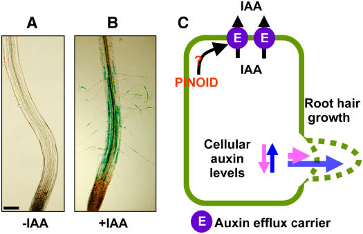
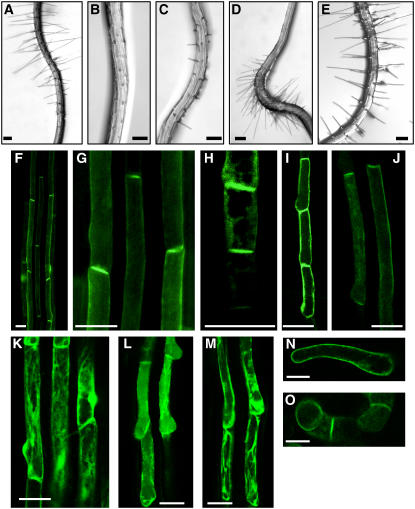
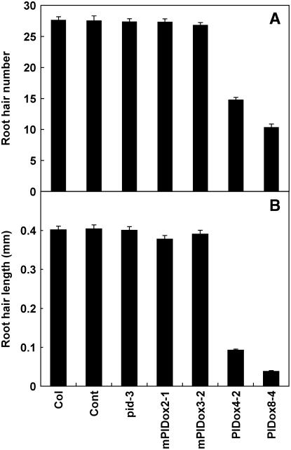
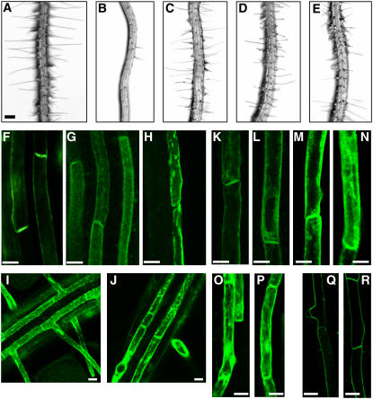
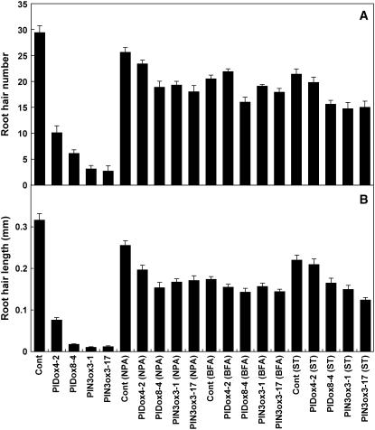
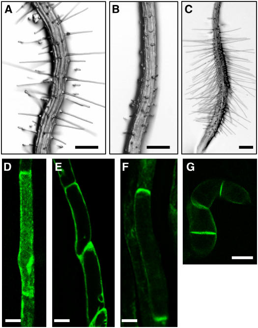
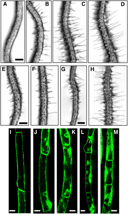
Intercellular transport of auxin is mediated by influx and efflux carriers in the plasma membrane and subjected to developmental and environmental regulation. Here, using the auxin-sensitive Arabidopsis thaliana root hair cell system and the tobacco (Nicotiana tabacum) suspension cell system, we demonstrate that the protein kinase PINOID (PID) positively regulates auxin efflux. Overexpression of PID (PIDox) or the auxin efflux carrier component PINFORMED3 (PIN3, PIN3ox), specifically in the root hair cell, greatly suppressed root hair growth. In both PIDox and PIN3ox transformants, root hair growth was nearly restored to wild-type levels by the addition of auxin, protein kinase inhibitors, or auxin efflux inhibitors. Localization of PID or PIN3 at the cell boundary was disrupted by brefeldin A and staurosporine. A mutation in the kinase domain abrogated the ability of PID to localize at the cell boundary and to inhibit root hair growth. These results suggest that PIDox- or PIN3ox-enhanced auxin efflux results in a shortage of intracellular auxin and a subsequent inhibition of root hair growth. In an auxin efflux assay using transgenic tobacco suspension cells, PIDox or PIN3ox also enhanced auxin efflux. Collectively, these results suggest that PID positively regulates cellular auxin efflux, most likely by modulating the trafficking of PIN and/or some other molecular partners involved in auxin efflux.


Figures

Figure 1.
Figure 1.
The Auxin-Sensitive Root Hair System and the Working Hypothesis of This Study. (A) and (B) Roots from hairless rhd6 mutant Arabidopsis harboring the PE7:GUS transgene. Seedlings were untreated (A) or treated with 30 nM IAA (B). IAA induces root hairs and hair cell–specific (PE7:GUS) expression in the transgenic mutant root. Bar = 100 μm. (C) Scheme of the regulation of intracellular auxin levels. Higher auxin efflux in the hair cell decreases cellular auxin levels and shortens the root hair. By contrast, less or normal auxin efflux activity maintains enough auxin inside the hair cell to sustain hair elongation. PID as a positive regulator of auxin efflux would inhibit root hair elongation if overexpressed.
Figure 2.
Figure 2.
Root Hair Cell–Specific Overexpression of PID Suppresses Root Hair Growth. (A) The control transformant (PE7:GFP) expressing only GFP in the hair cell, driven by the root hair cell–specific ATEXPA7 promoter (PE7). (B) to (D) PE7:PID-GFP transformants expressing PID-GFP in the hair cell, untreated ([B], severe phenotype transformant; [C], moderate phenotype transformant) or treated with 30 nM IAA (D). (E) PE7:mPID-GFP transformant expressing a mutated form of PID (mPID)–GFP with a point mutation in the ATP binding domain. (F) to (J) Confocal microscopy images of the roots of PE7:PID-GFP transformants. Images of fully elongated (or mature; [F], [G], [I], and [J]) and elongating (H) hair cells are shown. (K) A confocal microscopy image of the root of the control PE7:GFP transformant. (L) and (M) Confocal microscopy images of the roots of PE7:mPID-GFP transformants, expressing mPID-GFP. (N) and (O) Confocal microscopy images of tobacco BY-2 cells harboring PTA:PID-GFP. Cells were treated with dexamethasone (Dex) to induce expression, as described in Methods. Bars = 100 μm for (A) to (E) and 30 μm for (F) to (O).
Figure 3.
Figure 3.
Root Hair Development in PID Transformants. Root hair number (A) and root hair length (B) in control (Cont; PE7:GFP), loss-of-function pid-3 mutant, wild-type PID-overexpressing transformants (PE7:PID-GFP; PIDox), and mutant PID-overexpressing transformants (PE7:mPID-GFP; mPIDox). Two independent homozygous lines for each transformant were analyzed. Data represent means ± se for each line (n = 240 for root hair length, n = 30 for root hair number).
Figure 4.
Figure 4.
Effects of NPA, BFA, and ST on Root Hair Growth of PIDox Roots, and the Subcellular Localization of PID-GFP. (A) Control (PE7:GFP) transformant, untreated. Bar = 100 μm for (A) to (E). (B) to (E) Roots of PIDox (PE7:PID-GFP) transformants either untreated (B) or treated with 1 μM NPA (C), 5 μM BFA (D), or 0.25 μM ST (E). (F) to (H) Confocal microscopy images of roots from PE7:PID-GFP transformants treated with 0 μM (F), 10 μM (G), or 50 μM (H) BFA. Bars = 10 μm for (F) to (R). (I) and (J) Confocal microscopy images of roots of PE7:GFP transformants treated with 0 μM (I) or 50 μM (J) BFA. (K) to (N) Confocal microscopy images of root hair cells of PE7:PID-GFP transformants treated with 0 μM (K), 2 μM (L), or 5 μM ([M] and [N]) ST. (O) and (P) Confocal microscopy images of root hair cells of PE7:GFP transformants treated with 0 μM (O) or 5 μM (P) ST. (Q) and (R) Confocal microscopy images of root hair cells of PE7:AHA2-GFP transformants treated with 0 μM (Q) or 5 μM (R) ST.
Figure 5.
Figure 5.
NPA, BFA, and ST Restore Root Hair Growth of PIDox and PIN3ox Transformants. Root hair number (A) and root hair length (B) in control (Cont; PE7:GFP) transformants, PIDox transformants (PE7:PID-GFP), and PIN3ox transformants (PE7:PIN3-GFP) treated with 1 μM NPA, 5 μM BFA, or 0.25 μM ST. Two independent homozygous lines for each transformant were observed. Data represent means ± se for each transformant (n = 64 for root hair length, n = 8 for root hair number).
Figure 6.
Figure 6.
PID and PINs Are Expressed in the Root Hair Cell. RT-PCR analysis of RNA from Arabidopsis root hair cells (Root hair) or whole root tissues (Root). E7 (ATEXPA7) was used as a positive control for hair cell–specific amplification; GL2 (GLABRA2) was used as a control for non-hair cell–specific amplification. Gene-specific primer sets were designed to include intron(s) to discriminate amplification from cDNAs versus genomic DNA.
Figure 7.
Figure 7.
Hair Cell–Specific Overexpression of PIN3 Inhibits Root Hair Growth. (A) to (C) Roots from PE7:GFP (control; [A]) and PIN3ox (PE7:PIN3-GFP; [B] and [C]) transformants. Treatment with IAA (30 nM) restored root hairs in PIN3ox transformants (C). Bars = 100 μm. (D) to (F) Confocal microscopy images of the root hair cells from PE7:GFP (D) and PE7:PIN3-GFP ([E] and [F]) transformants. Bars = 10 μm. (G) A confocal microscopy image of transgenic tobacco BY-2 cells expressing PTA:PIN3-GFP. Bar = 30 μm.
Figure 8.
Figure 8.
Effects of NPA, BFA, and ST on Root Hair Growth of PIN3ox Roots, and Subcellular Localization of PIN3-GFP. (A) to (H) Roots of PIN3ox (PE7:PIN3-GFP) transformants treated with 0 μM (A), 0.1 μM (B), 0.5 μM (C), or 1.0 μM (D) NPA; 1.0 μM (E) or 5.0 μM (F) BFA; or 0.1 μM (G) or 0.5 μM (H) ST. Bars = 100 μm. (I) to (M) Confocal microscopy images of root hair cells from PE7:PIN3-GFP transformants. The seedlings were treated with 0 μM (I), 2 μM (J), or 5 μM (K) ST or 10 μM (L) or 50 μM (M) BFA. Bars = 10 μm.
Figure 9.
Figure 9.
Retention of [3H]NAA in Tobacco Cells Is Decreased by PIDox or PIN3ox. Relative retention of 3H-labeled NAA in PIDox (PTA:PID-GFP; [A]), PIN3ox (PTA:PIN3-GFP; [B]), and control (C) tobacco BY-2 cells. PID and PIN3 expression was driven by the Dex-inducible PTA promoter system for 24 h before measurement. Data represent means ± se from nine replicates. Averages for +Dex are significantly different from those for –Dex at P < 0.05 for (A) and P < 0.01 for (B).

References

    1. Aoyama, T., and Chua, N.-H. (1997). A glucocorticoid-mediated transcriptional induction system in transgenic plants. Plant J. 11 606–612. - PubMed
    1. Bechtold, N., and Pelletier, G. (1998). In planta Agrobacterium-mediated transformation of adult Arabidopsis thaliana plants by vacuum infiltration. In Arabidopsis Protocols, J.M. Martinez-Zapater and J. Salinas, eds (Totowa, NJ: Humana), pp. 259–266. - PubMed
    1. Becker, D., Kemper, E., Schell, J., and Masterson, R. (1992). New plant binary vectors with selectable markers located proximal to the left T-DNA border. Plant Mol. Biol. 20 1195–1197. - PubMed
    1. Benjamins, R., Quint, A., Weijers, D., Hooykaas, P., and Offringa, R. (2001). The PINOID protein kinase regulates organ development in Arabidopsis by enhancing polar auxin transport. Development 128 4057–4067. - PubMed
    1. Benková, E., Michniewicz, M., Sauer, M., Teichmann, T., Seifertová, D., Jürgens, G., and Friml, J. (2003). Local, efflux-dependent auxin gradients as a common module for plant organ formation. Cell 115 591–602. - PubMed

Publication types

MeSH terms