

My approach to the diagnosis of mesothelial lesions
- PMID: 16731600
- PMCID: PMC1860395
- DOI: 10.1136/jcp.2005.029652
My approach to the diagnosis of mesothelial lesions
Abstract
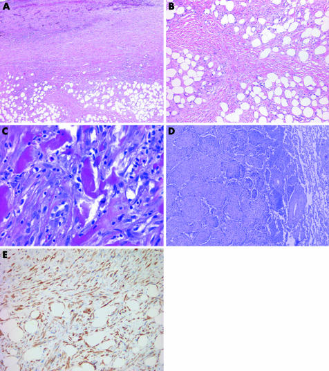
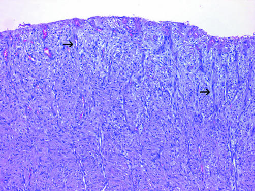
Mesothelial lesions pose considerable diagnostic challenges not only because benign tumours, reactive proliferations and malignant mesothelioma can mimic one another, but also because the morphological patterns displayed by malignant mesothelioma can simulate a variety of epithelial and non-epithelial malignancies. Immunohistochemical markers can aid in distinguishing epithelioid malignant mesothelioma from metastatic adenocarcinoma, but because no single marker reliably separates all cases, a panel of stains is recommended. Immunohistochemical studies are of more limited value in sarcomatoid malignant mesothelioma, and other features often play an essential role. The separation of reactive mesothelial proliferations from malignant mesothelioma on small biopsy can be quite difficult, as distinguishing features, such as stromal invasion, often cannot be adequately assessed. In adequately sampled lesions, however, the distinction between malignant mesothelioma, benign mesothelial proliferations and other tumours can be achieved in most cases by using a carefully integrated approach that incorporates clinical and radiographic data, immunohistochemical studies and, in selected cases, histochemical and ultrastructural techniques.
References
-
- Ordonez N G. The diagnostic utility of immunohistochemistry and electron microscopy in distinguishing between peritoneal mesotheliomas and serous carcinomas: a comparative study. Mod Pathol 20062696–100. - PubMed
-
- Baker P M, Clement P B, Young R H. Malignant peritoneal mesothelioma in women: a study of 75 cases with emphasis on their morphologic spectrum and differential diagnosis. Am J Clin Pathol 2005123724–737. - PubMed
-
- Ordonez N G. Value of cytokeratin 5/6 immunostaining in distinguishing epithelial mesothelioma of the pleura from lung adenocarcinoma. Am J Surg Pathol 1998221215–1221. - PubMed
-
- Chu P G, Weiss L M. Expression of cytokeratin 5/6 in epithelial neoplasms: an immunohistochemical study of 509 cases. Mod Pathol 2002156–10. - PubMed
MeSH terms
Substances
LinkOut - more resources
Full Text Sources
Medical