. 2006 Jun 6;103(23):8601-6.
doi: 10.1073/pnas.0511234103. Epub 2006 May 26.

Bacteriorhodopsin (bR) as an electronic conduction medium: current transport through bR-containing monolayers

Affiliations

Bacteriorhodopsin (bR) as an electronic conduction medium: current transport through bR-containing monolayers

Yongdong Jin et al. Proc Natl Acad Sci U S A. .

Abstract

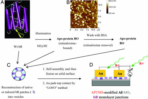
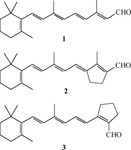
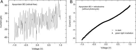
Studying electron transport (ET) through proteins is hampered by achieving reproducible experimental configurations, particularly electronic contacts to the proteins. The transmembrane protein bacteriorhodopsin (bR), a natural light-activated proton pump in purple membranes of Halobacterium salinarum, is well studied for biomolecular electronics because of its sturdiness over a wide range of conditions. To date, related studies of dry bR systems focused on photovoltage generation and photoconduction with multilayers, rather than on the ET ability of bR, which is understandable because ET across 5-nm-thick, apparently insulating membranes is not obvious. Here we show that electronic current passes through bR-containing artificial lipid bilayers in solid "electrode-bilayer-electrode" structures and that the current through the protein is more than four orders of magnitude higher than would be estimated for direct tunneling through 5-nm, water-free peptides. We find that ET occurs only if retinal or a close analogue is present in the protein. As long as the retinal can isomerize after light absorption, there is a photo-ET effect. The contribution of light-driven proton pumping to the steady-state photocurrents is negligible. Possible implications in view of the suggested early evolutionary origin of halobacteria are noted.


Conflict of interest statement

Conflict of interest statement: No conflicts declared.

Figures

Fig. 1.
Fig. 1.
Scheme of bR chemical tailoring and of the metal–protein–metal junction preparation. (A) Schematic representation of the 3D structure of bR. The seven α-helical domains form a transmembrane pore. The retinylidene residue is linked to the protein moiety via a protonated Schiff base linkage to Lys-216. (B) Representative AFM image (12 × 12 μm) of native bR patches prepared by 5 min of adsorption on an Al/AlOx substrate derivatized with APTMS. (C) Schematic of bR-containing vesicle. (D) Schematic of a Au/(single-bR-layer)/(APTMS-on-AlOx)/Al junction and measuring scheme.
Scheme 1.
Scheme 1.
Structures of all-trans retinal (native) (1), all-trans-locked (2), and 13-cis-locked (3) analogues.
Fig. 2.
Fig. 2.
AFM images of bR-containing fused membranes. (A Left) Representative AFM images (2 × 2 μm) of monolayers of fused membranes of bR-containing vesicles, prepared by 10-min adsorption on Al/AlOx substrate, derivatized with APTMS. (A Right and B Right) Line scans showing an average height of the strongest features of ≈5.2 nm. The height bar covers 20 nm. (B Left) A more densely packed bR containing a monolayer, prepared by 20-min adsorption of vesicles on the substrate (1.25- × 1.25-μm image). The crack in the membrane, induced by excessive drying (something that was carefully avoided in the preparation of the samples used for electrical transport measurements) shows the monolayer to be 5.1 nm thick (between markers).
Fig. 3.
Fig. 3.
IV characteristics of metal–(bR monolayer)–metal junctions. (A) IV curves of a Au/(wild-type bR)/(APTMS–AlOx–Al) junction containing oriented bR monolayer, prepared by vesicle fusion and measured at ambient conditions in the dark, upon illumination at λ > 550 nm, and dark adaptation. The arrows show that the IV responses can be cycled between the two states. The Au pad area was 2.10−3 cm2. (B) Plot of dark currents at a +1.0-V bias voltage for eight independent junctions prepared from bR/PC vesicles and from bR/OTG vesicles, respectively.
Fig. 4.
Fig. 4.
IV characteristics of metal–apo-membrane–metal junctions. (A) Typical IV characteristic of a Au/apo-membrane (retinal-free)/(APTMS–AlOx–Al) junction prepared by vesicle fusion and measured at ambient conditions. (B) IV curves of Au/apo-membrane (apo-protein bR plus retinaloxime)/(APTMS–AlOx–Al) junction prepared by vesicle fusion and measured at ambient conditions in the dark and upon illumination at λ > 550 nm. No photoeffect on the junction current was observed.

References

    1. Oesterhelt D., Stoeckenius W. Nat. New Biol. 1971;233:149–152. - PubMed
    1. Henderson R., Unwin P. N. T. Nature. 1975;257:28–32. - PubMed
    1. Lanyi J. K. Mol. Membr. Biol. 2004;21:143–150. - PubMed
    1. Pettei M. J., Yudd A. P., Nakanishi K., Henselman R., Stoeckenius W. Biochemistry. 1977;16:1955–1959. - PubMed
    1. Birge R. R., Gillespie N. B., Izaguirre E. W., Kusnetzow A., Lawrence A. F., Singh D., Song Q. W., Schmidt E., Stuart J. A., Seetharaman S., Wise K. J. J. Phys. Chem. B. 1999;103:10746–10766.

Publication types

MeSH terms