. 2006 Jun 6;103(23):8709-14.
doi: 10.1073/pnas.0508006103. Epub 2006 May 26.

Accumulation of small murine minor satellite transcripts leads to impaired centromeric architecture and function

Affiliations

Accumulation of small murine minor satellite transcripts leads to impaired centromeric architecture and function

Haniaa Bouzinba-Segard et al. Proc Natl Acad Sci U S A. .

Abstract

RNAs have been implicated in the assembly and stabilization of large-scale chromatin structures including centromeric architecture; unidentified RNAs are integral components of human pericentric heterochromatin and are required for localization of the heterochromatin protein HP1 to centromeric regions. Because satellite repeats in centromeric regions are known to be transcribed, we assessed a role for noncoding centromeric RNAs in the structure and function of the centromere. We identified minor satellite transcripts of 120 nt in murine cells that localize to centromeres and accumulate upon stress or differentiation. Forced accumulation of 120-nt transcripts leads to defects in chromosome segregation and sister-chromatid cohesion, changes in hallmark centromeric epigenetic markers, and mislocalization of centromere-associated proteins essential for centromere function. These findings suggest that small centromeric RNAs may represent one of many pathways that regulate heterochromatin assembly in mammals, possibly through tethering of kinetochore- and heterochromatin-associated proteins.


Conflict of interest statement

Conflict of interest statement: No conflicts declared.

Figures
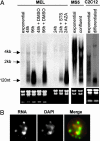
Fig. 1.
Fig. 1.
The major detectable form of murine satellite repeats transcripts is 120-nt long. (A) Total RNA extracts from murine cell lines or primary cells were analyzed by Northern blot to determine the size and relative abundance of transcripts from centromeric minor satellite repeats in exponentially growing, confluent, or differentiated cells. The minor satellite repeat unit fragment, in the sense orientation, was used as a probe. The profile of ribosomal 28S, 18S, and 5S, stained with ethidium bromide, is shown below each blot to confirm equal loading of the lanes. Cell lines analyzed were MEL in exponentially growing phase or after 48 and 96 h in culture, with or without the inducer of differentiation (DMSO), or MEL cells treated for 24 h with the inhibitor of DNA methylation 5-azacytidine (AZA) or the inducer of apoptosis staurosporine (STS) (Left); murine primary stromal cells, exponentially growing or confluent myoblast cells (Center, MS5), and exponentially growing or differentiated myoblast cells (Right, C2C12). (B) RNA-FISH analysis on confluent MEL cells, using a biotinylated probe identical to the one used in Northern blots. (Left) Centromeric transcripts. (Center) DAPI. (Right) Merge of both images, with RNA in green and DAPI in red.
Fig. 2.
Fig. 2.
Ectopic expression of satellite transcripts leads to impaired mitosis and lack of sister-chromatid cohesion. (A) Examples of interphase and mitotic nuclei from pSAT-transfected cells after cell sorting. Cells were fixed and nuclei stained with DAPI. The images show normal mitotic figures in GFP cells (Left, arrows) and accumulation of mitosis with misaligned chromosomes in pSAT/GFP+-transfected cells (Right, arrows). (B) Examples of DAPI-stained chromosomes from metaphase spreads. (Left) GFP showing normal chromosome shape and chromatid cohesion. (Right) pSAT/GFP+-transfected MEL showing impaired sister-chromatid cohesion and centromere cleavage (arrows).
Fig. 3.
Fig. 3.
Accumulation of centromeric transcripts leads to mislocalization of centromere-associated proteins. Immunofluorescence on pSAT-transfected MEL cells [GFP cells (Left) and GFP+ cells (Right)] using antibodies against (at left, from bottom to top) the chromosomal passenger protein Aurora-B, the centromere-associated protein HP1γ, or the histone H3 trimethylated on K9 (H3MeK9) and MafK. Chromosomes are stained with DAPI (Center). Specific staining appears in red and chromosomes in blue in the merge lanes.

References

    1. Choo K. H. The Centromere. New York: Oxford Univ. Press; 1997.
    1. Henikoff S., Ahmad K., Platero J. S., van Steensel B. Proc. Natl. Acad. Sci. USA. 2000;97:716–721. - PMC - PubMed
    1. Vafa O., Sullivan K. F. Curr. Biol. 1997;7:897–900. - PubMed
    1. Guenatri M., Bailly D., Maison C., Almouzni G. J. Cell Biol. 2004;166:493–505. - PMC - PubMed
    1. Sullivan B. A., Karpen G. H. Nat. Struct. Mol. Biol. 2004;11:1076–1083. - PMC - PubMed

Publication types