. 2006 Jun;80(12):5686-96.
doi: 10.1128/JVI.02739-05.

Sindbis virus nonstructural protein nsP2 is cytotoxic and inhibits cellular transcription

Affiliations

Sindbis virus nonstructural protein nsP2 is cytotoxic and inhibits cellular transcription

Natalia Garmashova et al. J Virol. 2006 Jun.

Abstract

Replication of alphaviruses in vertebrate cells strongly affects cell physiology and ultimately leads to development of a cytopathic effect (CPE) and cell death. Sindbis virus (SIN) replication causes major changes in cellular macromolecular synthesis, in which the strong downregulation of transcription of cellular mRNAs and rRNAs plays a critical role. SIN nonstructural protein nsP2 was previously proposed as one of the main regulators of virus-host cell interactions, because point mutations in the carboxy-terminal part of nsP2 could make SIN and other alphaviruses and replicons less cytopathic and capable of persisting in some vertebrate cell lines. These mutants were incapable of inhibiting transcription and downregulating a viral stress-induced cell response. In the present work, we demonstrate that (i) SIN nsP2 is critically involved in CPE development, not only during the replication of SIN-specific RNAs, but also when this protein is expressed alone from different expression cassettes; (ii) the cytotoxic effect of SIN nsP2 appears to be at least partially determined by its ability to cause transcriptional shutoff; (iii) these functions of SIN nsP2 are determined by the integrity of the carboxy-terminal peptide of this protein located outside its helicase and protease domains, rather than by its protease activity; and (iv) the cytotoxic activity of SIN nsP2 depends on the presence of this protein in a free form, and alterations in P123 processing abolish the ability of nsP2 to cause CPE.


Figures

FIG. 1.
FIG. 1.
Analysis of the effect of SIN nsP2 expression on cell survival. (A) Schematic representation of VEE replicons expressing SIN nsP2 and analysis of their ability to establish persistent replication. Arrows indicate the positions of the subgenomic promoters. Ub indicates an ubiquitine sequence fused in frame with the SIN nsP2 gene. Pac indicates the puromycin acetyltransferase gene. Different dilutions of the electroporated cells were seeded into 100-mm tissue culture dishes. Puromycin selection was performed as described in Materials and Methods. Purr cell colonies were stained with crystal violet at days 4 to 9 posttransfection, depending on their growth rates. The results are presented in CFU per μg of RNA used for transfection. The ranges indicate variations between the experiments. (B) Analysis of replicon genome RNA replication and transcription of the subgenomic RNAs. The details of the experiments are described in Materials and Methods. RNA labeling was performed with [3H]uridine between 15 and 20 h postelectroporation. The positions of genomic and subgenomic RNAs are indicated. (C and D) Analysis of growth of the cells transfected with VEE replicons expressing GFP (C), and analysis of cell survival at different times posttransfection with VEE replicons expressing wt SIN nsP2 (D). Equal numbers of cells were seeded into six-well Costar plates (one-tenth of the electroporated cells per well). Puromycin selection (10 μg/ml) was performed between 6 and 48 h posttransfection. The cells were then incubated in puromycin-free medium, and viable cells were counted at the indicated times. The data were normalized on the number of viable adherent cells determined at 6 h posttransfection. Panels C and D present the results of the same experiment. The data were separated between two panels for clarity of presentation, because the cells containing GFP-expressing replicons were capable of efficient growth, and cells with VEE replicons expressing wt SIN nsP2 were dying. One of the multiple reproducible experiments is presented. The standard deviations are indicated. (E) Accumulation of SIN nsP2 in the BHK-21 cells transfected with VEE replicons expressing wt SIN nsP2 and with SINrep/GFP replicon (8). Cell lysates were prepared at 20 h posttransfection and analyzed by Western blotting as described in Materials and Methods.
FIG. 2.
FIG. 2.
Accumulation of mutations in wt SIN nsP2 encoded by VEE replicons. Purr cell colonies formed after transfection of VEErep/nsP2/Pac and VEErepL/nsP2/Pac replicons were randomly selected, and the genome fragment encoding SIN nsP2 was sequenced. Replicons in some of the clones expressed multiple variants of the SIN nsP2-coding gene.
FIG. 3.
FIG. 3.
Analysis of the effect of mutated SIN nsP2 on cytotoxicity of the VEE replicons. (A) Schematic representation of VEE genome-based replicons expressing SIN nsP2 containing the previously identified adaptive mutations (9) and analysis of their ability to establish persistent replication and develop Purr foci. Arrows indicate the positions of the subgenomic promoters. Ub indicates a ubiquitine sequence fused in frame with the SIN nsP2 gene. (B) Analysis of growth of the cells carrying VEE replicons expressing SIN nsP2 with P726→G and P726→L mutations. The details of the experimental procedure are described in Materials and Methods.
FIG. 4.
FIG. 4.
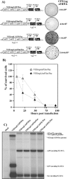
Analysis of the cytotoxicity of SIN nsP2 with mutated protease. (A) Schematic representation of VEE genome-based replicons expressing SIN nsP2 containing C481→S mutation and analysis of their ability to establish persistent replication and develop Purr foci. (B) Survival of the cells transfected with VEE replicons expressing SIN nsP2 with C481→S mutation. The data were normalized on the number of viable adherent cells determined at 6 h posttransfection. (C) Analysis of replicons' genome replication and transcription of the subgenomic RNAs. RNA labeling was performed with [32P]phosphoric acid. The details of the experiments are described in Materials and Methods.
FIG. 5.
FIG. 5.
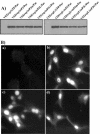
Accumulation of SIN nsP2 in the BHK-21 cells transfected with VEE replicons expressing different forms of SIN nsP2. (A) Cell lysates were prepared at 20 h posttransfection and analyzed by Western blotting as described in Materials and Methods. (B) For analysis of nsP2 distribution, BHK-21 cells were electroporated by in vitro-synthesized RNAs and, at 16 h posttransfection, stained with rabbit anti-SIN nsP2 and goat anti-rabbit IgG Alexa Fluor 546-labeled secondary antibodies (Molecular Probes). Cells: a, VEErepL/GFP/Pac-transfected cells; b, VEErepL/nsP2/Pac-transfected cells; c, VEErepL/nsP2L/Pac-transfected cells; d, VEErepL/nsP2m/Pac-transfected cells.
FIG. 6.
FIG. 6.
Inhibition of transcription and development of apoptosis in the BHK-21 cells transfected with VEE replicons expressing different forms of SIN nsP2. (A) BHK-21 cells were electroporated by 5 μg of the in vitro-synthesized RNAs. At 15 h posttransfection, cellular RNAs were labeled with [3H]uridine for 5 h and analyzed by RNA gel electrophoresis under the conditions described in Materials and Methods. For quantitative analysis, the rRNA bands were excised from the PPO-impregnated gels (shown in the upper panel), and the radioactivity was measured by liquid scintillation counting (lower panel). One of two reproducible experiments is presented. (B) For detection of apoptosis, BHK-21 cells were electroporated by in vitro-synthesized RNAs and, at 48 h posttransfection, stained with mouse anti-phospho-histone H2A.X antibodies and goat anti-mouse IgG Alexa Fluor 546-labeled secondary antibodies (Molecular Probes) as described in Materials and Methods in subpanels b, d, f, h, and j. DAPI staining of the same cells is shown in subpanels a, c, e, g, and i. Cells: a and b, VEErepL/GFP/Pac-transfected cells; c and d, VEErepL/nsP2/Pac-transfected cells; e and f, VEErepL/nsP2L/Pac-transfected cells; g and h, VEErepL/nsP2G/Pac-transfected cells; i and j, VEErepL/nsP2m/Pac-transfected cells.
FIG. 7.
FIG. 7.
Analysis of the effect of SIN nsP2 expression from plasmid DNAs on cell survival. (A) Schematic representation of plasmid-based cassettes, containing chicken β-actin promoter and expressing different forms of SIN nsP2. Ubi indicates a ubiquitine sequence fused in frame with SIN nsP2 gene. (B and C) Analysis of the cell survival (B) and cell growth (C) at different times posttransfection with the plasmids. The details of the experimental procedure are given in Materials and Methods. Panels B and C present the results of the same experiment. The data were separated between two panels for clarity of presentation. The data were normalized on the number of viable adherent cells determined at 24 h posttransfection.

Similar articles

Cited by

References

    1. Agapov, E. V., I. Frolov, B. D. Lindenbach, B. M. Prägai, S. Schlesinger, and C. M. Rice. 1998. Noncytopathic Sindbis virus RNA vectors for heterologous gene expression. Proc. Natl. Acad. Sci. USA 95:12989-12994. - PMC - PubMed
    1. Basler, C. F., X. Wang, E. Muhlberger, V. Volchkov, J. Paragas, H. D. Klenk, A. Garcia-Sastre, and P. Palese. 2000. The Ebola virus VP35 protein functions as a type I IFN antagonist. Proc. Natl. Acad. Sci. USA 97:12289-12294. - PMC - PubMed
    1. Billecocq, A., M. Spiegel, P. Vialat, A. Kohl, F. Weber, M. Bouloy, and O. Haller. 2004. NSs protein of Rift Valley fever virus blocks interferon production by inhibiting host gene transcription. J. Virol. 78:9798-9806. - PMC - PubMed
    1. Bouloy, M., C. Janzen, P. Vialat, H. Khun, J. Pavlovic, M. Huerre, and O. Haller. 2001. Genetic evidence for an interferon-antagonistic function of rift valley fever virus nonstructural protein NSs. J. Virol. 75:1371-1377. - PMC - PubMed
    1. Bredenbeek, P. J., I. Frolov, C. M. Rice, and S. Schlesinger. 1993. Sindbis virus expression vectors: packaging of RNA replicons by using defective helper RNAs. J. Virol. 67:6439-6446. - PMC - PubMed

Publication types