. 2006 Jun;80(12):5976-83.
doi: 10.1128/JVI.00110-06.

Properties and dissemination of H5N1 viruses isolated during an influenza outbreak in migratory waterfowl in western China

Affiliations

Properties and dissemination of H5N1 viruses isolated during an influenza outbreak in migratory waterfowl in western China

Hualan Chen et al. J Virol. 2006 Jun.

Abstract

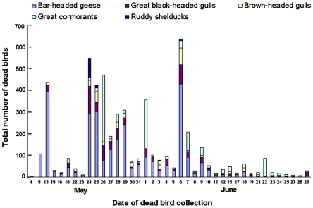
H5N1 influenza A viruses are widely distributed among poultry in Asia, but until recently, only a limited number of wild birds were affected. During late April through June 2005, an outbreak of H5N1 virus infection occurred among wild birds at Qinghai Lake in China. Here, we describe the features of this outbreak. First identified in bar-headed geese, the disease soon spread to other avian species populating the lake. Sequence analysis of 15 viruses representing six avian species and collected at different times during the outbreak revealed four different H5N1 genotypes. Most of the isolates possessed lysine at position 627 in the PB2 protein, a residue known to be associated with virulence in mice and adaptation to humans. However, neither of the two index viruses possessed this residue. All of the viruses tested were pathogenic in mice, with the exception of one index virus. We also tested the replication of two viruses isolated during the Qinghai Lake outbreak and one unrelated duck H5N1 virus in rhesus macaques. The Qinghai Lake viruses did not replicate efficiently in these animals, producing no evidence of disease other than transient fever, while the duck virus replicated in multiple organs and caused symptoms of respiratory illness. Importantly, H5N1 viruses isolated in Mongolia, Russia, Inner Mongolia, and the Liaoning Province of China after August 2005 were genetically closely related to one of the genotypes isolated during the Qinghai outbreak, suggesting the dominant nature of this genotype and underscoring the need for worldwide intensive surveillance to minimize its devastating consequences.


Figures

FIG. 1.
FIG. 1.
Course of migratory waterfowl deaths due to H5N1 viruses at Qinghai Lake. A total of 6,184 dead birds were collected from 4 May to 29 June 2005: 3,282 bar-headed geese, 929 great black-headed gulls, 570 brown-headed gulls, 1,302 great cormorants, and 145 ruddy shelducks.
FIG. 2.
FIG. 2.
Pattern of spread of H5N1 viruses of different genotypes among wild birds at Qinghai Lake. The disease began in bar-headed geese, spread to brown-headed gulls and great black-headed gulls, and then spread to great cormorants and ruddy shelducks. The viruses from these species represent four genotypes, two of which appear to have spread from bar-headed geese to the other three avian species, although it is uncertain whether the genotype D virus originated in the bar-headed goose population.
FIG. 3.
FIG. 3.
Histopathologic analysis of a moribund bar-headed goose from the H5N1 virus outbreak at Qinghai Lake. (a) Brain showing scattered nonsuppurative inflammatory foci characterized by perivascular cuffing of mononuclear cells, microgliosis, degeneration of nerve cells, and edema (HE stain). (b) Brain with numerous nerve and glial cells positive for viral antigen (brown pigments) by immunohistological staining with an anti-H5 polyclonal antibody. (c) Pancreas showing scattered coagulative necrotic foci in parenchyma with nonsuppurative inflammation (HE stain). (d) Heart showing small nonsuppurative inflammatory foci with degenerating cardiomyocytes (HE stain).
FIG. 4.
FIG. 4.
Phylogenetic analyses of the H5N1 viruses isolated during the Qinghai Lake outbreak. The phylogenetic trees were generated with the PHYLIP program of the CLUSTALX software package (version 1.81) by using the neighbor-joining algorithm and bootstrap values of 1,000. (a) HA (nucleotides 105 to 1659); (b) PB2 (nucleotides 82 to 2264); (c) PA (nucleotides 67 to 2151). The phylogenetic tree of HA was rooted to A/mallard/Denmark/64650/03 (H5N7), and the PB2 and PA phylogenetic trees were rooted to A/Memphis/1/90 (H3N2). The sequences of the wild-bird viruses obtained in this study are shown in red, and the viruses isolated after the Qinghai Lake outbreak are shown in green, while those of the wild-bird viruses reported previously by Liu et al. and Chen et al. are shown in blue. Dates of virus isolation during Qinghai Lake outbreak are also shown.
FIG. 5.
FIG. 5.
Histological findings from rhesus macaques infected with H5N1 viruses. (a) Section from a consolidated area from lungs shows bronchointerstitial pneumonia with severe infiltration of inflammatory cells (BHGs/QH/1/05 virus, day 7 postinfection) (HE stain). The lung lesions were distributed around the bronchioli. Asterisks indicate lumen of bronchioli. (b) Severe alveolar damage was observed within and along the periphery of the consolidated area (BHGs/QH/1/05 virus, day 7 postinfection) (HE stain). Severe proliferative and reactive hyperplasia of alveolar cells with massive recruitment of lymphocytes, fibrin exudates, and alveolar edema are shown. (c) A strong reaction with macrophages was one of the prominent findings in the lungs (BHGs/QH/1/05 virus, day 7 postinfection) (HE stain). (d) Severe alveolar edema, thickening of alveolar wall with lymphocyte recruitment (white arrow), and regeneration of alveolar cells (black arrow) were also observed (BHGs/QH/1/05 virus, day 7 postinfection) (HE stain). (e) The lung lesions were detected as peribronchiolitis in a macaque infected with DK/GX/35/01 virus at 4 days postinfection (DK/GX/35/01 virus, day 4 postinfection) (HE stain). The asterisk indicates lumen of bronchioles. (f) Prominent alveolar edema and strong reaction with foamy macrophages but scant regenerative change and scant lymphocytic recruitment in a macaque infected with DK/GX/35/01 virus (DK/GX/35/01 virus, day 7 postinfection) (HE stain). (g) Viral antigens in tonsilar epithelium on day 4 postinfection (brown) (BHGs/QH/1/05 virus, day 4 postinfection) (immunohistochemistry).

References

    1. Chen, H., G. Deng, Z. Li, G. Tian, Y. Li, P. Jiao, L. Zhang, Z. Liu, R. G. Webster, and K. Yu. 2004. The evolution of H5N1 influenza viruses in ducks in southern China. Proc. Natl. Acad. Sci. USA 101:10452-10457. - PMC - PubMed
    1. Chen, H., G. J. D. Smith, S. Y. Zhang, K. Qin, J. Wang, K. S. Li, R. G. Webster, J. S. M. Peiris, and Y. Guan. 2005. Avian flu: H5N1 virus outbreak in migratory waterfowl. Nature 436:191-192. - PubMed
    1. Claas, E. C., A. D. M. E. Osterhaus, R. van Beek, J. C. De Jong, G. F. Rimmelzwaan, D. A. Senne, S. Krauss, K. F. Shortridge, and R. G. Webster. 1998. Human influenza A H5N1 virus related to a highly pathogenic avian influenza virus. Lancet 351:472-477. - PubMed
    1. Clements, J. F. 2000. Birds of the world: a checklist. Ibis, Vista, Calif.
    1. Fouchier, R., P. Schneeberger, F. Rozendaal, J. Broekman, S. Kemink, V. Munster, T. Kuiken, G. Rimmelzwaan, M. Schutten, G. van Doornum, K. Guus, B. Arnold, K. Marion, and A. D. M. E. Osterhaus. 2004. Avian influenza A virus (H7N7) associated with human conjunctivitis and a fatal case of acute respiratory distress syndrome. Proc. Natl. Acad. Sci. USA 101:1356-1361. - PMC - PubMed

Publication types