. 2006 Jun;80(12):6072-83.
doi: 10.1128/JVI.02495-05.

Dissociation of a MAVS/IPS-1/VISA/Cardif-IKKepsilon molecular complex from the mitochondrial outer membrane by hepatitis C virus NS3-4A proteolytic cleavage

Affiliations

Dissociation of a MAVS/IPS-1/VISA/Cardif-IKKepsilon molecular complex from the mitochondrial outer membrane by hepatitis C virus NS3-4A proteolytic cleavage

Rongtuan Lin et al. J Virol. 2006 Jun.

Abstract

Intracellular RNA virus infection is detected by the cytoplasmic RNA helicase RIG-I that plays an essential role in signaling to the host antiviral response. Recently, the adapter molecule that links RIG-I sensing of incoming viral RNA to downstream signaling and gene activation events was characterized by four different groups; MAVS/IPS-1-1/VISA/Cardif contains an amino-terminal CARD domain and a carboxyl-terminal mitochondrial transmembrane sequence that localizes to the mitochondrial membrane. Furthermore, the hepatitis C virus NS3-4A protease complex specifically targets MAVS/IPS-1/VISA/Cardif for cleavage as part of its immune evasion strategy. With a novel search program written in python, we also identified an uncharacterized protein, KIAA1271 (K1271), containing a single CARD-like domain at the N terminus and a Leu-Val-rich C terminus that is identical to that of MAVS/IPS-1/VISA/Cardif. Using a combination of biochemical analysis, subcellular fractionation, and confocal microscopy, we now demonstrate that NS3-4A cleavage of MAVS/IPS-1/VISA/Cardif/K1271 results in its dissociation from the mitochondrial membrane and disrupts signaling to the antiviral immune response. Furthermore, virus-induced IKKepsilon kinase, but not TBK1, colocalized strongly with MAVS at the mitochondrial membrane, and the localization of both molecules was disrupted by NS3-4A expression. Mutation of the critical cysteine 508 to alanine was sufficient to maintain mitochondrial localization of MAVS/IPS-1/VISA/Cardif and IKKepsilon in the presence of NS3-4A. These observations provide an outline of the mechanism by which hepatitis C virus evades the interferon antiviral response.


Figures
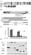
FIG. 1.
FIG. 1.
Characterization of the RIG-I adapter. (A) Alignment of the CARD domains of Mda-5, RIG-I, and K1271. A BLAST alignment reveals CARD domain homology among the N-terminal sequences of RIG-I, Mda-5, and K1271. C-terminal alignment of the Leu-Val-rich region of K1271 and Bcl-xL is also shown. (B) Schematic representation of K1271. The 540-aa MAVS/IPS-1/VISA/Cardif molecule is shown schematically. The location of the CARD domain-containing, proline-rich region that interacts with TRAF6 and the C-terminal mitochondrial membrane region (TM) is shown. Also illustrated is the region adjacent to the TM containing cysteine 508, the target residues for the HCV NS3-4A protease. The different N- and C-terminal deletions used in this study are shown below the schematic. (C) N- and C-terminal domains of K1271 are required for IFN-β promoter activation. HEK293 cells were transfected with 100 ng of pRLTK control plasmid and 100 ng of IFN-β-pGL3 reporter plasmid, with increasing amounts (200 ng and 1,000 ng) of different truncated forms of Myc-K1271 expression constructs as indicated. Luciferase activity was analyzed at 24 h posttransfection by the Dual-Luciferase reporter assay as described by the manufacturer (Promega). Relative luciferase activity was measured as activation (n-fold; relative to the basal level of reporter gene in the presence of pcDNA3 vector after normalization with cotransfected renilla luciferase activity); values are means ± standard deviations from three experiments. (D) Localization of active forms of K1271 to the insoluble fraction of the cytoplasm. The soluble lysates and insoluble fractions were prepared from the HEK293 cells used for panel C, equilibrated to the same volumes by sodium dodecyl sulfate-polyacrylamide gel electrophoresis loading buffer, and analyzed by immunoblotting with anti-Myc antibody 9E10.
FIG. 2.
FIG. 2.
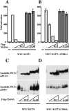
Cleavage of K1271 and inhibition of K1271-mediated transactivation by NS3-4A. (A) HEK293 cells were transfected with 100 ng of pRLTK control plasmid, 100 ng of IFN-β-pGL3 reporter plasmid, and 200 ng of K1271 (A) or K1271(A508) (B) Myc-tagged expression plasmid, together with increasing amounts (250 ng, 500 ng, and 1,000 ng) of protease-active NS3-4A or inactive NS3-4A(A139) Flag-tagged expression plasmid as indicated. Luciferase activity was analyzed at 24 h posttransfection. (C and D) The soluble lysates and insoluble fractions were prepared from HEK293 cells used for panels A and B, equilibrated to the same volumes by sodium dodecyl sulfate-polyacrylamide gel electrophoresis loading buffer, and analyzed by immunoblotting with anti-Flag antibody M2 and anti-Myc antibody 9E10.
FIG. 3.
FIG. 3.
Localization of K1271 to the mitochondria. HEK293 cells were transfected with combinations of 200 ng of IKKɛ and RIG-I; in some cases, cells were also infected with Sendai virus (40 hemagglutinating units/ml) at 24 h posttransfection for a total of 12 h. Mitochondrial extracts were prepared, and 50 μg of extract was run on 7.5% sodium dodecyl sulfate-polyacrylamide gel electrophoresis and immunoblotted with anti-K1271 antibody.
FIG. 4.
FIG. 4.
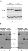
Endogenous K1271 is cleaved in Huh8 HCV replicon cells. (A) Whole-cell extracts (50 μg) prepared from uninfected or Sendai virus-infected Huh7 or Huh8 cells were subjected to sodium dodecyl sulfate-polyacrylamide gel electrophoresis (SDS-PAGE) and probed with anti-K1271 and antiactin antibodies. (B) Mitochondrial and cytosolic fractions (50 μg) from HEK293T cells transfected with 200 ng of NS3-4A expression plasmid (lanes 1 to 4) were subjected to SDS-PAGE and analyzed by immunoblotting with anti-K1271, antitubulin, anti-cytochrome c, and anti-hsp70. Mitochondrial and cytosolic fractions (50 μg) from Huh7 (H7) and Huh8 (H8) cells (lanes 5 to 8) were also subjected to SDS-PAGE and analyzed by immunoblotting with anti-K1271, antitubulin, anti-hsp70, and anti-cytochrome c.
FIG. 5.
FIG. 5.
Endogenous K1271 is cleaved in Huh8 HCV replicon cells. Huh7 (A to C) and Huh8 (D to F) cells were treated with MTO (B and E), fixed in paraformaldehyde, and stained for K1271 (A and D). Confocal fluorescent images were merged (C and F). K1271 was detected using rabbit anti-K1271 C-term and anti-rabbit AF488 antibodies.
FIG. 6.
FIG. 6.
Colocalization of endogenous IKKɛ and K1271. Human lung epithelial A549 cells, either control (upper panels) or VSV infected (lower panels), were costained for IKKɛ (in green) and K1271 (in red), and confocal fluorescent images were merged. IKKɛ was detected using mouse anti-IKKɛ and anti-mouse AF488 antibodies, while K1271 was detected using guinea pig anti-K1271 N-term and anti-guinea pig AF546 antibodies.
FIG. 7.
FIG. 7.
Distinct subcellular localization of IKKɛ and TBK1. VSV-infected A549 cells were treated with MTO (in red), fixed, and stained for either IKKɛ (upper panels) or TBK1 (lower panels). The confocal fluorescent images were merged. IKKɛ was detected as described above, while TBK1 was detected using rabbit anti-TBK1 and anti-rabbit AF488 antibodies.
FIG. 8.
FIG. 8.
Disruption of the IKKɛ-K1271 complex localization by expression of NS3-4A. COS-7 cells transfected with IKKɛ and K1271 expression plasmids, without (upper panels) or with (lower panels) a construct encoding NS3-4A, were costained for IKKɛ (in green) and K1271 (in red), and confocal fluorescent images were merged. IKKɛ and K1271 were detected as described above.
FIG. 9.
FIG. 9.
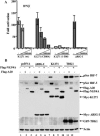
Inhibition of IRF and IFN-β activation by NS3-4A. (A) Inhibition of the IFN-β promoter. HEK293 cells were transfected with 100 ng of pRLTK control plasmid, 100 ng of IFN-β-pGL3 reporter plasmid, and 200 ng of ΔRIG-I and wild-type or mutated forms of K1271 expression plasmids, together with increasing amounts of NS3-4A expression plasmid (125 ng, 500 ng, and 2,000 ng) as indicated. Luciferase activity was analyzed at 24 h posttransfection by the Dual-Luciferase reporter assay as described by the manufacturer (Promega). Relative luciferase activity was measured as activation (n-fold; relative to the basal level of reporter gene in the presence of pcDNA3 vector after normalization with cotransfected renilla luciferase activity); values are means ± standard deviations from three experiments. (B) A20 and NS3-4A inhibit K1271- and ΔRIG-I-mediated activation of IRF-3 and IRF-7. HEK293 cells were cotransfected with 1 μg of IRF-7, IRF-3, Myc-ΔRIG-I, Myc-K1271, or GFP-TBK1 and 2 μg of Flag-A20 or Flag-NS3-4A expression construct as indicated. Whole-cell extracts (50 μg) were resolved by sodium dodecyl sulfate-polyacrylamide gel electrophoresis and analyzed by immunoblotting for IRF-7 pSer477/479, IRF-3 pSer396, Flag-A20, Flag-NS3-4A, Myc-ΔRIG-I, Myc-K1271, GFP-TBK1, and actin.
FIG. 10.
FIG. 10.
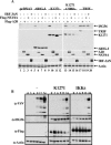
(A) NS3-4A and A20 inhibit K1271-mediated activation of endogenous ISG56 gene expression. HEK293 cells were cotransfected with 1 μg of pcDNA3, Myc-ΔRIG-I, Myc-KIAA1271, Myc-KIAA1271(C508A), or Myc-TRIF and 2 μg of IRF-3ΔN, Flag-A20, or Flag-NS3-4A expression construct as indicated. Whole-cell extracts (50 μg) were resolved by sodium dodecyl sulfate-polyacrylamide gel electrophoresis and analyzed by immunoblotting using antibodies to ISG56, Myc, Flag, IRF-3, and actin. (B) K1271 inhibits VSV replication. HEK293 cell lines expressing K1271 or IKKɛ were infected with VSV (1 multiplicity of infection), and viral protein expression was measured at different times after infection by immunoblotting. Expression of the Flag-tagged transgene and the endogenous ISG56 was also monitored.

Similar articles

Cited by

References

    1. Akira, S., and K. Takeda. 2004. Toll-like receptor signalling. Nat. Rev. Immunol. 4:499-511. - PubMed
    1. Alexopoulou, L., A. C. Holt, R. Medzhitov, and R. A. Flavell. 2001. Recognition of double-stranded RNA and activation of NF-kappaB by Toll-like receptor 3. Nature 413:732-738. - PubMed
    1. Alter, H. J., and L. B. Seeff. 2000. Recovery, persistence, and sequelae in hepatitis C virus infection: a perspective on long-term outcome. Semin. Liver Dis. 20:17-35. - PubMed
    1. Andrejeva, J., K. S. Childs, D. F. Young, T. S. Carlos, N. Stock, S. Goodbourn, and R. E. Randall. 2004. The V proteins of paramyxoviruses bind the IFN-inducible RNA helicase, mda-5, and inhibit its activation of the IFN-beta promoter. Proc. Natl. Acad. Sci. USA 101:17264-17269. - PMC - PubMed
    1. Blight, K. J., A. A. Kolykhalov, and C. M. Rice. 2000. Efficient initiation of HCV RNA replication in cell culture. Science 290:1972-1974. - PubMed

Publication types

MeSH terms

LinkOut - more resources