. 2006 Jun;15(6):1239-47.
doi: 10.1110/ps.062076806.

Structure of the 21-30 fragment of amyloid beta-protein

Affiliations

Structure of the 21-30 fragment of amyloid beta-protein

Andrij Baumketner et al. Protein Sci. 2006 Jun.

Abstract

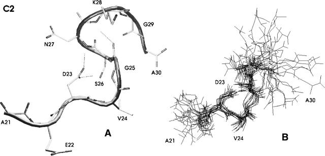
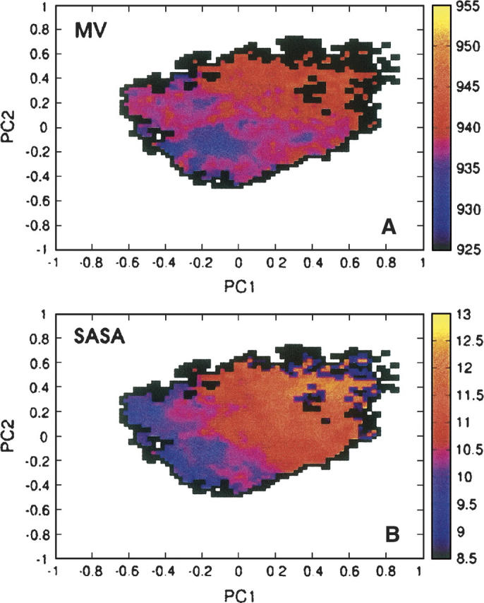
Folding and self-assembly of the 42-residue amyloid beta-protein (Abeta) are linked to Alzheimer's disease (AD). The 21-30 region of Abeta, Abeta(21-30), is resistant to proteolysis and is believed to nucleate the folding of full-length Abeta. The conformational space accessible to the Abeta(21-30) peptide is investigated by using replica exchange molecular dynamics simulations in explicit solvent. Conformations belonging to the global free energy minimum (the "native" state) from simulation are in good agreement with reported NMR structures. These conformations possess a bend motif spanning the central residues V24-K28. This bend is stabilized by a network of hydrogen bonds involving the side chain of residue D23 and the amide hydrogens of adjacent residues G25, S26, N27, and K28, as well as by a salt bridge formed between side chains of K28 and E22. The non-native states of this peptide are compact and retain a native-like bend topology. The persistence of structure in the denatured state may account for the resistance of this peptide to protease degradation and aggregation, even at elevated temperatures.


Figures

Figure 1.
Figure 1.
Conformational ensembles from replica exchange molecular dynamics simulations of Aβ(21–30). (A) Free energy (in units of kT) as a function of the first two principal components PC1 and PC2 at T = 300 K. (B) Projections of the 10 most populated conformational clusters C1–C10 onto PC1–PC2 space. The most frequently visited cluster C1 is ∼30% populated.
Figure 2.
Figure 2.
The most populated structure observed in replica exchange simulations of Aβ(21–30). (A) Centroid conformation of the cluster (cluster C1) and (B) a number of cluster members superimposed onto each other to demonstrate the extent of structural variability.
Figure 3.
Figure 3.
Formation of salt bridges in the most populated cluster C1 between the positively-charged side chain of K28 and a number of negatively-charged residues and the C terminus. Histograms are shown for distances between K28:Nζ and E22:Cδ, K28:Nζ and D23:Cγ, and K28:Nζ and A10:C. Data for the K28–E22 pair reveal both close contact and solvent-mediated salt bridges.
Figure 4.
Figure 4.
The second most-populated structure observed in replica exchange simulations of Aβ(21–30). (A) Centroid conformation of the cluster (cluster C2) and (B) a number of cluster members superimposed onto each other to demonstrate the extent of structural variability.
Figure 5.
Figure 5.
Mean molecular volume (in units of Å3) (A) and solvent-accessible surface area (in units of nm2) (B), as a function of the two first principal components PC1,PC2 generated for conformations sampled in the replica exchange simulations.
Figure 6.
Figure 6.
Secondary structure assignments for the individual amino acid residues of Aβ(21–30), determined by using the DSSP algorithm (Kabsch and Sander 1983). The assignments—(A-Helix) α–helix; (3-Helix) 310-helix; (Beta-S) β-sheet; (Beta-B) β–bridge; (Bend) bends; (Turns) β–turns; (Coil) all other conformations—are shown separately for the most populated cluster C1 (“native” state) (A), and all other conformations sampled in the simulations (“denatured” state) (B).

Similar articles

Cited by

References

    1. Antzutkin O.N., Leapman R.D., Balbach J.J., Tycko R. 2002. Supramolecular structural constraints on Alzheimer's β-amyloid fibrils from electron microscopy and solid-state nuclear magnetic resonance Biochemistry 41 15436–15450. - PubMed
    1. Berendsen H.J.C., van der Spoel D., van Drunen R. 1995. GROMACS: A message-passing parallel molecular dynamics implementation Comput. Phys. Commun. 91 43–56.
    1. Borreguero J.M., Urbanc B., Lazo N.D., Buldyrev S.V., Teplow D.B., Stanley H.E. 2005. Folding events in the 21-30 region of amyloid β-protein (Aβ) studied in silico Prot. Natl. Acad. Sci. 102 6015–6020. - PMC - PubMed
    1. Bryngelson J.D. and Wolynes P.G. 1987. Spin glasses and the statistical mechanics of protein folding Proc. Natl. Acad. Sci. 84 7524–7528. - PMC - PubMed
    1. Chimon S. and Ishii Y. 2005. Capturing intermediate structures of Alzheimer's β-amyloid, Aβ (1-40), by solid-state NMR spectroscopy J. Am. Chem. Soc. 127 13472–13473. - PubMed

Publication types

MeSH terms