. 2006 Jun;15(6):1248-59.
doi: 10.1110/ps.051995006.

Solution structure of the ubiquitin-associated domain of human BMSC-UbP and its complex with ubiquitin

Affiliations

Solution structure of the ubiquitin-associated domain of human BMSC-UbP and its complex with ubiquitin

Yong-Gang Chang et al. Protein Sci. 2006 Jun.

Abstract

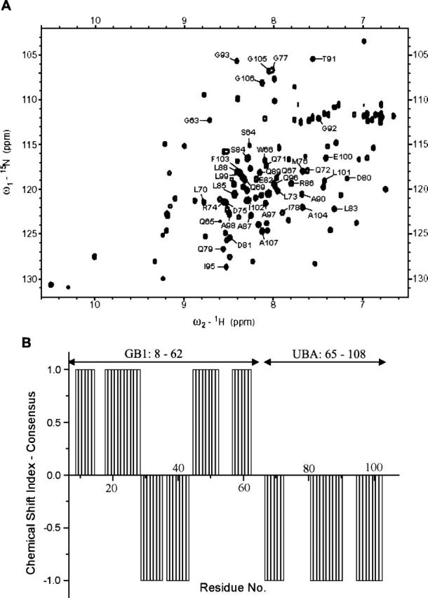
Ubiquitin is an important cellular signal that targets proteins for degradation or regulates their functions. The previously identified BMSC-UbP protein derived from bone marrow stromal cells contains a ubiquitin-associated (UBA) domain at the C terminus that has been implicated in linking cellular processes and the ubiquitin system. Here, we report the solution NMR structure of the UBA domain of human BMSC-UbP protein and its complex with ubiquitin. The structure determination was facilitated by using a solubility-enhancement tag (SET) GB1, immunoglobulin G binding domain 1 of Streptococcal protein G. The results show that BMSC-UbP UBA domain is primarily comprised of three alpha-helices with a hydrophobic patch defined by residues within the C terminus of helix-1, loop-1, and helix-3. The M-G-I motif is similar to the M/L-G-F/Y motifs conserved in most UBA domains. Chemical shift perturbation study revealed that the UBA domain binds with the conserved five-stranded beta-sheet of ubiquitin via hydrophobic interactions with the dissociation constant (KD) of approximately 17 microM. The structural model of BMSC-UbP UBA domain complexed with ubiquitin was constructed by chemical shift mapping combined with the program HADDOCK, which is in agreement with the result from mutagenesis studies. In the complex structure, three residues (Met76, Ile78, and Leu99) of BMSC-UbP UBA form a trident anchoring the domain to the hydrophobic concave surface of ubiquitin defined by residues Leu8, Ile44, His68, and Val70. This complex structure may provide clues for BMSC-UbP functions and structural insights into the UBA domains of other ubiquitin-associated proteins that share high sequence homology with BMSC-UbP UBA domain.


Figures

Figure 1.
Figure 1.
Multiple alignments of selected UBA domains. (A) Schematic representation of several UBA-containing proteins showing that UBA domains usually locate at the C terminus, whereas the accompanying UBQ domains occur at the N terminus. UBA domains are in black, and UBQ domains are in gray. Residue numbers are shown to the right of the diagram of each protein. Hu, Homo sapiens. (B) Sequence alignment of BMSC-UbP_UBA (338–380), p62_UBA (387–440), HHR23A_UBA(1) (163–200), and HHR23A_UBA(2) (320–363) whose structures have been determined by NMR. Amino acid residues in gray indicate identity or similarity. (C) Sequence alignment of the UBA domains of ubiquilin-1 (also called hPLIC1), ubiquilin-2 (hPLIC2), A1Up and yeast Dsk2, showing that they share high sequence homology. The SwissProt entry codes for the sequences are Q96S82 (BMSC-UbP), P48510 (Dsk2), Q9UMX0 (ubiquilin-1), Q9UHD9 (ubiquilin-2), Q9NRR5 (A1Up), P54725 (HHR23A), and Q13501 (p62).
Figure 2.
Figure 2.
Sequence-specific assignment and secondary structure identification for the HGB1-UBA fusion protein. (A) 1H-15N HSQC spectrum of HGB1-UBA. Both GB1 and the UBA domain have wide chemical-shift dispersions in the spectrum. Almost all the 1H-15N correlation peaks have been assigned by heteronuclear multidimensional NMR techniques. Only the peaks attributed to the backbone amides of BMSC-UbP UBA domain (residues 65–108) are labeled in the figure. (B) Chemical shift index (CSI) reflecting the secondary structures of HGB1-UBA. CSI consensus shows the identification of secondary structures using the chemical shifts of 1Hα, 13Cα, 13Cβ, and 13CO of HGB1-UBA. The indices indicate residues residing in α-helix (−1) and β-sheet (+1), indicative of three α-helix composition of the UBA domain under study. The GB1 (residues 8–62) and UBA (65–108) domains are indicated, respectively, while the N-terminal His6-tag and two additional residues (Gly63 and Ser64) from the BamHI restriction site are omitted.
Figure 3.
Figure 3.
Solution structure of BMSC-UbP UBA domain. (A) Superimposition of the backbone Cα traces representing a bundle of 10 refined structures. (B) Ribbon representation of the structure of BMSC-UbP UBA domain. The three helices from the N to the C terminus are labeled as α1, α2, and α3. (C) Structure of BMSC-UbP UBA domain showing the characteristic hydrophobic core. The side chains of the residues that contribute to the hydrophobic core are depicted in neon style.
Figure 4.
Figure 4.
Comparison of the UBA domain structures. Shown are the ribbon representations of BMSC-UbP UBA (A), HHR23A UBA(2) (B), p62 UBA (C), and HHR23A UBA(1) (D). The side chains of the residues in the conserved M/L-G-F/Y/I motifs are highlighted in neon style. The PDB codes for the structures are 2CWB (BMSC-UbP UBA), 1DV0 [HHR23A UBA(2)], 1QO2 (p62 UBA), and 1IFY [HHR23A UBA(1)], respectively.
Figure 5.
Figure 5.
Determination of the binding affinity of BMSC-UbP UBA for ubiquitin. (A) Overlay of the 1H-15N HSQC spectra of ubiquitin free (red) and bound (black) to BMSC-UbP UBA. The molar ratio of ubiquitin to HGB1-UBA is 1:4. The three lines indicate the typical chemical-shift changes of residues Ile13, Lys48, and Leu71 of 15N-labeled ubiquitin upon titrating with HGB1-UBA. (B) Plot of weighted average chemical shift changes (Δδave) for Ile13 (▪), Lys48 (•), and Leu71 (▴) against the molar ratio of HGB1-UBA:ubiquitin. The curves indicate the most favorable fitting using the equation described in the Materials and Methods section.
Figure 6.
Figure 6.
Mapping the contact surface of BMSC-UbP UBA and ubiquitin by chemical shift perturbation. (A) Chemical shift changes of HGB1-UBA vs. its residue number. 15N-labeled HGB1-UBA was titrated stepwise with unlabeled ubiquitin to a final molar ratio of 1:4. The average chemical shift change (Δδave) for every amide is plotted against residue number of HGB1-UBA. The solid and the dashed lines indicate the threshold values of mean and mean + SD for the chemical shift changes. Residues with the chemical shift changes above the mean + SD (dashed line) are considered involved in significant contact with ubiquitin as shown in B. (B) Mapping the significant contact residues on the BMSC-UbP UBA structure, including Trp66 (helix1); Met76, Gly77 and Ile78 (loop 1); Ala98, Leu99, Glu100 and L101 (helix 3). The side chains of these residues are highlighted in neon style. (C) As in A, except for the titration of 15N-labeled ubiquitin with HGB1-UBA. (D) As in B. The residues with significant chemical shift change are Leu8 and Ile13 (loop between β1 and β2), Leu43 (β3), Ala46 (loop between β3 and β4), Lys48 and Gln49 (β4), Leu71 (β5), and Arg72 (C terminus). The ubiquitin structure is referenced from the crystal structure (PDB code 1UBQ).
Figure 7.
Figure 7.
Structure of BMSC-UbP UBA-ubiquitin complex. (A) Superimposition of the backbone Cα traces representing 10 refined structures of BMSC-UbP UBA–ubiquitin complex. Ubiquitin is shown in blue; BMSC-UbP UBA, in green. (B) Ribbon representation of the complex structure. (C) Interaction surfaces of BMSC-UbP UBA and ubiquitin. Electrostatic potential maps are shown for ubiquitin. BMSC-UbP UBA is represented in round ribbon except for omission of α2 for clarity. Side chains of residues Met76, Ile78, and Leu99 are shown in green and in neon style as indicated by red arrows. (D) Side chains of the hydrophobic residues that are in close contact: Met76, Ile78, and Leu99 of BMSC-UbP UBA (green) and Leu8, Ile44, His68, and Val70 of ubiquitin (bright gray) as displayed in neon style. The side chains of residues Lys48 and Lys63 are shown in blue, which face away from the contact surface in the complex despite large chemical shift change of Lys48 upon titration. (E) Overlay of the UBA domains from BMSC-UbP (residues 68–104, green) and Dsk2 (334–370, red) in each UBA–ubiquitin complex. (F) Structure of yeast Dsk2 UBA–ubiquitin complex for comparison. The ubiquitin molecule is positioned in the same orientation as that in B.
Figure 8.
Figure 8.
Mutations reveal the specific binding surfaces on BMSC-UbP UBA and ubiquitin. (A) Plot of weighted average chemical shift changes (Δδave) of Leu71 of 15N-labeled ubiquitin against the molar ratio of HGB1-UBA mutants (M76A, I78A, L99A, and L101A) to ubiquitin. By data analysis, the dissociation constants of M76A and I78A are >200 μM, while those of L99A and L101A are 51 and 12 μM, respectively. (B) The average chemical shift changes of Glu100 of 15N-labeled HGB1-UBA vs. the molar ratio of ubiquitin and K48A mutant to HGB1-UBA. The dissociation constant of K48A binding with HGB1-UBA is 9 μM, which is comparable with that of wild-type ubiquitin binding (∼5 μM). The curves indicate the most favorable fitting using the equation described in the Materials and Methods section.

References

    1. Alam S.L., Sun J., Payne M., Welch B.D., Blake B.K., Davis D.R., Meyer H.H., Emr S.D., Sundquist W.I. 2004. Ubiquitin interactions of NZF zinc fingers EMBO J. 23 1411–1421. - PMC - PubMed
    1. Beal R., Deveraux Q., Xia G., Rechsteiner M., Pickart C. 1996. Surface hydrophobic residues of multiubiquitin chains essential for proteolytic targeting Proc. Natl. Acad. Sci. 93 861–866. - PMC - PubMed
    1. Bertolaet B.L., Clarke D.J., Wolff M., Watson M.H., Henze M., Divita G., Reed S.I. 2001. UBA domains mediate protein-protein interactions between two DNA damage-inducible proteins J. Mol. Biol. 313 955–963. - PubMed
    1. Chen L., Shinde U., Ortolan T.G., Madura K. 2001. Ubiquitin-associated (UBA) domains in Rad23 bind ubiquitin and promote inhibition of multi-ubiquitin chain assembly EMBO Rep. 2 933–938. - PMC - PubMed
    1. Ciani B., Layfield R., Cavey J.R., Sheppard P.W., Searle M.S. 2003. Structure of the ubiquitin-associated domain of p62 (SQSTM1) and implications for mutations that cause Paget's disease of bone J. Biol. Chem. 278 37409–37412. - PubMed

Publication types

MeSH terms

Associated data

LinkOut - more resources