Clinical Trial
. 2007 Feb;56(2):155-63.
doi: 10.1007/s00262-006-0181-3. Epub 2006 May 30.

In vivo tracking of macrophage activated killer cells to sites of metastatic ovarian carcinoma

Affiliations
Clinical Trial

In vivo tracking of macrophage activated killer cells to sites of metastatic ovarian carcinoma

D Ritchie et al. Cancer Immunol Immunother. 2007 Feb.

Abstract

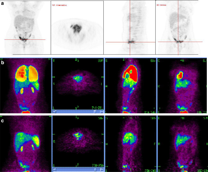
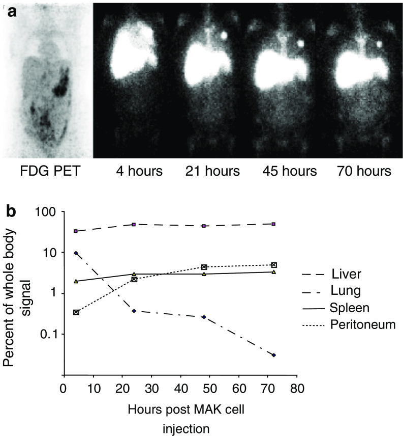
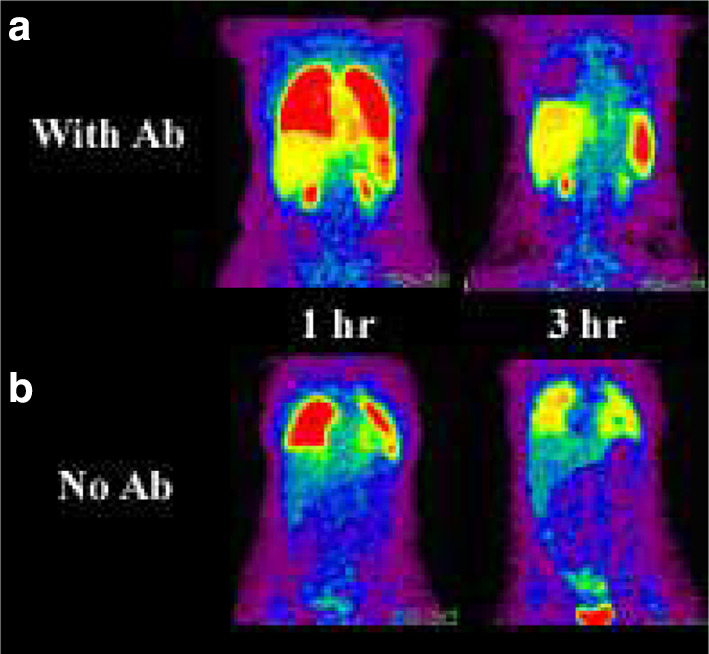
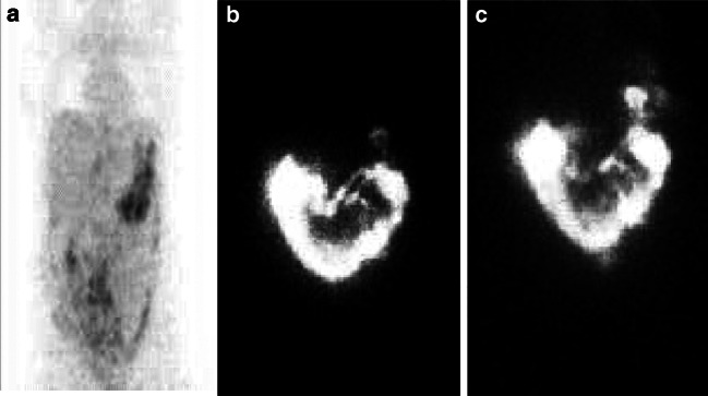
Radio-labelling of blood cells is an established technique for evaluating in vivo migration of normal cells to sites of pathology such as infection and haemorrhage. A limitation of cellular immunotherapies to induce anti-tumour responses is in part due to the uncertain ability of cellular effectors to reach their intended target. We extended the approach of cell radiolabelling to accurately examine the in vivo distribution of cellular immunotherapy with ex-vivo macrophage activated killer (MAK) cells. We describe the use of two methods of cell labelling for tracking the destination of autologous-derived macrophage activated killer (MAK) cells linked to the bi-specific antibody MDX-H210 delivered either by intravenous (i.v.) or intraperitoneal (i.p.) injection in ten patients with peritoneal relapse of epithelial ovarian carcinoma. Our results demonstrate the feasibility of generating high numbers and purity of GMP quality MAK cells, which can be radiolabelled with (18)F-FDG or (111)In-oxime. MAK cell administration produced minimal infusional toxicity and demonstrated a reproducible pattern of in vivo distribution and active in vivo tracking to sites of known tumour following 8 of 16 i.v. infusions or 4 of 6 i.p. infusions. However, the leakage of (18)F-FDG limited the ability to confidently confirm the tracking of MAK cells to tumour in all cases and improved PET labels are required. The addition of MDX-H210 bispecific antibody did not alter the distribution of cells to tumour sites, but did accelerate the clearance of i.v. administered MAK cells from the pulmonary circulation. This data demonstrates that cellular cancer immunotherapies may be successfully delivered to the sites of active tumour following either i.v. or i.p. injection in a proportion of patients with metastatic cancer. Incorporation of tracking studies in early cycles of cellular immunotherapy may allow selection of patients who demonstrate successful targeting of the immunotherapy for ongoing treatment.


Figures

Fig. 1
Fig. 1
Baseline and serial PET images in patient 7 after i.v. injection of MAK cells labelled with18F-FDG+ MDX-H210 antibody. a Baseline PET images obtained on a combined PET/CT scanner 2 weeks prior to MAK cell injection following direct intravenous administration of 111MBq 18F-FDG demonstrating multi-focal peritoneal metastases predominantly in the pelvis and additional lesions in the serosal peritoneum over the liver and anterior superior tip of the spleen. b Scans 1 h after injection: whole body projection image (left), and orthogonal axial through pelvis (second from left), sagittal (second from right) and coronal (right) planes demonstrate MAK cells predominantly within the lungs with early uptake in liver, spleen and pelvic tumor. c Scans 4 h after injection: whole body images (left), axial through pelvis (second from left), sagittal (second from right) and coronal (right) demonstrate activity predominantly within the liver, spleen and localized to sites of pelvic peritoneal tumor deposits
Fig. 2
Fig. 2
a Baseline PET images and serial planar whole body imaging in patient 4 after i.v. injection of 111indium labelled MAK cells + MDX-H210 antibody. Far left panel: baseline PET images demonstrating mildly increased radiotracer uptake through out the peritoneal cavity with nodular areas of moderately increased radiotracer uptake in the left upper quadrant, right iliac fossa and the presacral space consistent with peritoneal deposits. Other upper panels: serial whole body images of 111indium labelled MAK cells at 4, 21, 45 and 70 h post-injection. At 4 h, diffuse activity was seen throughout both lung fields, consistent with normal white cell margination. Three foci of moderately intense activity in the left lung were consistent with micro-emboli of radiolabelled cells to this lung. At 21 h there was progressive clearance of activity from the lungs and increasing activity in the liver, spleen and bone marrow. At 21 h there was faint visualization of the peritoneal space. This became more obvious by 70 h where there was clearly a diffuse pattern of uptake in the abdomen with most marked uptake in the left mid abdomen corresponding to the area of most marked abnormality on the baseline 18F-FDG scan. b Distribution of injected MAK cells by organ of interest in patient 4. Signals present in each organ at each time point are expressed as a percentage of the whole body signal at that time. Data presented are typical for that obtained in six separate administrations
Fig. 3
Fig. 3
Comparison of 18F-FDG-labelled MAK cell biodistribution with (a) and without (b) antibody. Images obtained at 1 h after reinfusion of radiolabelled cells (left panels) demonstrates significantly greater liver, splenic and renal activity when antibody is attached to the cell. This likely reflects more rapid demargination of cells from the pulmonary vasculature. By 3 h the antibody-labelled cells had almost completely cleared from the lungs whereas the cells without antibody remained to a greater extent marginated in the lungs with relatively less liver and particularly splenic localization.There is a reciprocal earlier appearance of antibody labelled MAK cells within the liver and spleen and in the areas of known metastatic tumor in the right iliac fossa
Fig. 4
Fig. 4
Progressive accumulation of 111In-labelled MAK cells + MDX-H210 antibody in the left upper quadrant area of known metastatic tumor in patient 4. a A pre-MAK cell administration PET scan reveals radiotracer uptake in the left upper quadrant of the abdomen. b Following i.p. administration of In-labelled MAK cells intraperitoneal distribution throughout the peritoneum is seen with early and then progressive tracking of MAK cells to the known tumor site at 3.5 h. c Progressive tracking observed at 18 h

References

    1. Ribas A, Butterfield LH, Glaspy JA, Economou JS. Current developments in cancer vaccines and cellular immunotherapy. J Clin Oncol. 2003;21:2415–2432. doi: 10.1200/JCO.2003.06.041. - DOI - PubMed
    1. Frangioni J, Hajjar R. In vivo tracking of stem cells for clinical trials in cardiovascular disease. Circulation. 2004;110:3378–3384. doi: 10.1161/01.CIR.0000149840.46523.FC. - DOI - PubMed
    1. Chokri M, Lopez M, Oleron C, Girard A, Martinache C, Siffert JC, Canepa S, Bartholeyns J. Production of macrophages with potent antitumoral properties (MAK) by culture of monocytes in the presence of GM-CSF and 1,25 (OH)2 vitD3. Anticancer Res. 1992;12:2257–2260. - PubMed
    1. Munn D, Cheung N. Phagocytosis of tumor cells by human monocytes cultured in GM-CSF. J Exp Med. 1990;172:231–237. doi: 10.1084/jem.172.1.231. - DOI - PMC - PubMed
    1. Fidler I, Kleinerman E. Therapy of cancer metastases by systemic activation of macrophages. Res Immunol. 1992;144:284–287. doi: 10.1016/0923-2494(93)80109-C. - DOI - PubMed

Publication types

MeSH terms