

Biexponential characterization of prostate tissue water diffusion decay curves over an extended b-factor range
- PMID: 16735177
- PMCID: PMC1880900
- DOI: 10.1016/j.mri.2005.12.008
Biexponential characterization of prostate tissue water diffusion decay curves over an extended b-factor range
Abstract
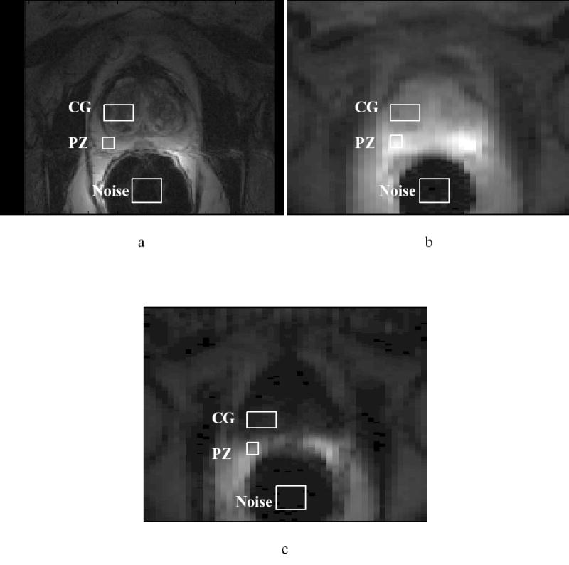
Detailed measurements of water diffusion within the prostate over an extended b-factor range were performed to assess whether the standard assumption of monoexponential signal decay is appropriate in this organ. From nine men undergoing prostate MR staging examinations at 1.5 T, a single 10-mm-thick axial slice was scanned with a line scan diffusion imaging sequence in which 14 equally spaced b factors from 5 to 3,500 s/mm(2) were sampled along three orthogonal diffusion sensitization directions in 6 min. Due to the combination of long scan time and limited volume coverage associated with the multi-b-factor, multidirectional sampling, the slice was chosen online from the available T2-weighted axial images with the specific goal of enabling the sampling of presumed noncancerous regions of interest (ROIs) within the central gland (CG) and peripheral zone (PZ). Histology from prescan biopsy (n=9) and postsurgical resection (n=4) was subsequently employed to help confirm that the ROIs sampled were noncancerous. The CG ROIs were characterized from the T2-weighted images as primarily mixtures of glandular and stromal benign prostatic hyperplasia, which is prevalent in this population. The water signal decays with b factor from all ROIs were clearly non-monoexponential and better served with bi- vs. monoexponential fits, as tested using chi(2)-based F test analyses. Fits to biexponential decay functions yielded intersubject fast diffusion component fractions in the order of 0.73+/-0.08 for both CG and PZ ROIs, fast diffusion coefficients of 2.68+/-0.39 and 2.52+/-0.38 microm(2)/ms and slow diffusion coefficients of 0.44+/-0.16 and 0.23+/-0.16 um(2)/ms for CG and PZ ROIs, respectively. The difference between the slow diffusion coefficients within CG and PZ was statistically significant as assessed with a Mann-Whitney nonparametric test (P<.05). We conclude that a monoexponential model for water diffusion decay in prostate tissue is inadequate when a large range of b factors is sampled and that biexponential analyses are better suited for characterizing prostate diffusion decay curves.
Figures

References
-
- American Cancer Society. Cancer Facts and Figures. 2004.
-
- Heuck A, Scheidler J, Sommer B, Graser A, Muller-Lisse UG, Massmann J. MR imaging of prostate cancer. Radiologue. 2003;43:464–473. - PubMed
-
- Mueller-Lisse UG, Mueller-Lisse UL, Haller S, Schneede P, Scheidler JE, Schmeller N, Hofstetter AG, Reiser MF. Likelihood of prostate cancer based on prostate-specific antigen density by MRI: Retrospective analysis. JCAT. 2002;26:432–437. - PubMed
-
- Tuzel E, Sevinc M, Obuz F, Sade M, Kirkali Z. Is magnetic resonance imaging necessary in the staging of prostate cancer? Urol Internat. 1998;61:227–231. - PubMed
-
- Perrotti M, Han KR, Epstein RE, Kennedy EC, Rabbani F, Badani K, Pantuck AJ, Weiss RE, Cummings KB. Prospective evaluation of endorectal magnetic resonance imaging to detect tumor foci in men with prior negative prostatic biopsy: A pilot study. J Urol. 1999;162:1314–1317. - PubMed
Publication types
MeSH terms
Grants and funding
- R03 HS013234/HS/AHRQ HHS/United States
- R03HS13234-01/HS/AHRQ HHS/United States
- U41 RR019703/RR/NCRR NIH HHS/United States
- 1R01CA109246-01A/CA/NCI NIH HHS/United States
- AG P01CA67165-03/AG/NIA NIH HHS/United States
- 1 R33 CA99015/CA/NCI NIH HHS/United States
- R01 AG19513-01/AG/NIA NIH HHS/United States
- R01 AG019513/AG/NIA NIH HHS/United States
- R25 CA089017/CA/NCI NIH HHS/United States
- 1 R01 NS39335-01A1/NS/NINDS NIH HHS/United States
- R01 CA109246/CA/NCI NIH HHS/United States
- R01 NS039335/NS/NINDS NIH HHS/United States
- P01 CA067165/CA/NCI NIH HHS/United States
LinkOut - more resources
Full Text Sources
Other Literature Sources
Medical
