. 2006 Jun 6;103(23):8864-9.
doi: 10.1073/pnas.0603214103. Epub 2006 May 30.

Polymerization of hyperphosphorylated tau into filaments eliminates its inhibitory activity

Affiliations

Polymerization of hyperphosphorylated tau into filaments eliminates its inhibitory activity

Alejandra del C Alonso et al. Proc Natl Acad Sci U S A. .

Abstract

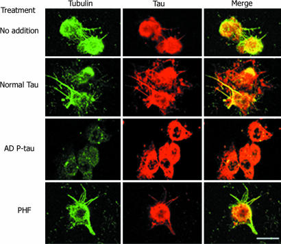
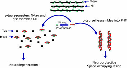
Accumulation of abnormally hyperphosphorylated tau (P-tau) in the form of tangles of paired helical filaments and/or straight filaments is one of the hallmarks of Alzheimer's disease (AD) and other tauopathies. P-tau is also found unpolymerized in AD. Although the cognitive decline is known to correlate with the degree of neurofibrillary pathology, whether the formation of filaments or the preceding abnormal hyperphosphorylation of tau is the inhibitory entity that leads to neurodegeneration has been elusive. We have previously shown that cytosolic abnormally hyperphosphorylated tau in AD brain (AD P-tau) sequesters normal tau (N-tau), microtubule-associated protein (MAP) 1, and MAP2, which results in the inhibition of microtubule assembly and disruption of microtubules. Here, we show that polymerization of AD P-tau into filaments inhibits its ability to bind N-tau and as well as the ability to inhibit the assembly of tubulin into microtubules in vitro and in the regenerating microtubule system from cultured cells. Like AD P-tau, the in vitro abnormally hyperphosphorylated recombinant brain N-tau binds N-tau and loses this binding activity on polymerization into filaments. Dissociation of the hyperphosphorylated N-tau filaments by ultrasonication restores its ability to bind N-tau. These findings suggest that the nonfibrillized P-tau is most likely the responsible entity for the disruption of microtubules in neurons in AD. The efforts in finding a therapeutic intervention for tau-induced neurodegeneration need to be directed either to prevent the abnormal hyperphosphorylation of this protein or to neutralize its binding to normal MAPs, rather than to prevent its aggregation into filaments.


Conflict of interest statement

Conflict of interest statement: No conflicts declared.

Figures

Fig. 1
Fig. 1
Coomassie blue staining, Western blotting, and the binding of N-tau to AD P-tau, PHF, and in vitro hyperphosphorylated N-tau. (A) N-tau (50 ng) and 4-μg aliquots of AD P-tau and PHF were subjected to SDS/PAGE, transferred to poly(vinylidene difluoride) membrane, and immunodetected with Tau-1 Ab. One strip was stained with Coomassie blue (Cb). Before immunostaining, the blots were treated with (+) or without (−) alkaline phosphatase (AP, 100 units/ml) for 3 h at 35°C. (B) Increasing concentrations of AD P-tau and duplicates of three different preparations of PHF, and as a control N-tau (10–100 μg tau content), were spotted on nitrocellulose membrane and overlaid with 5 μg/ml τ4L or with BSA and developed with Tau-1 Ab. To verify that the proteins bound to the membrane, one strip without any overlay was developed with a mixture of three phospho-independent polyclonal Abs to total tau. (C) Quantitations of the tau bound to AD P-tau (■) and to PHF (▴). (D) N-tau was hyperphosphorylated in vitro, as described in ref. ; one aliquot was probe-sonicated for 1 min to disrupt the tau filaments formed during the incubation period (19). The sonicated sample was centrifuged at 55,000 × g for 20 min, and the pellet was discarded. The nonsonicated and the supernatant of the sonicated samples were dotted in triplicates on three nitrocellulose membrane strips. These strips were processed as described above. The quantitations are given as mean ± SD. ∗, P < 0.005.
Fig. 2
Fig. 2
Effect of AD P-tau and PHF on microtubule assembly. Inhibition of microtubule assembly was studied as described in Materials and Methods. The assembly reaction was carried out by using 2 mg/ml tubulin mixed with 0.1 mg/ml N-tau alone (curve 1), with 0.2 mg/ml AD P-tau (curve 2), with 0.2 mg/ml self-assembled AD P-tau (curve 3), or 0.2 mg/ml PHF (curve 4). Tubulin without any addition was incubated as a control (curve 5). AD P-tau inhibited the microtubule-assembly-promoting activity of N-tau (compare curves 1 and 2), whereas PHF did not show any significant effect (compare curves 1 and 4 as well as 1 and 3, respectively).
Fig. 3
Fig. 3
AD P-tau but not PHF inhibits regeneration of microtubules from 3T3 cells. The extracted cells were incubated with 15% fresh rat brain cytosol in buffer containing 1 mM GTP for 1 h at 37°C, as described in Materials and Methods. Cells were processed for immunofluorescence staining by using DM1-A Ab against tubulin (green) and 134d rabbit polyclonal Ab against tau (red). Only AD P-tau could inhibit microtubule assembly.
Fig. 4
Fig. 4
Effect of AD P-tau self-assembly on its binding to N-tau. Self-assembly of AD P-tau was induced as described in Materials and Methods. At 0, 60, and 120 min of its self-assembly, aliquots were both examined by negative stained electron microscopy and spotted on nitrocellulose membrane, and overlaid with 5 μg/ml τ4L to determine the binding of N-tau, as described in Fig. 1B. At time 0 min, AD P-tau was nonfibrillized (B), and, after 120 min, bundles of PHF (C) could be detected by electron microscopy. The nonfibrillized AD P-tau bound N-tau and the amount of tau bound decreased with the degree of AD P-tau polymerization (A). (Inset) Nitrocellulose membrane spotted with AD P-tau at different time periods of incubation during the polymerization reaction, then overlaid with nothing, BSA, or N-tau and developed with Tau-1 or Abs against total tau.
Fig. 5
Fig. 5
Proposed mechanism of tau-induced neurodegeneration in AD and related tauopathies.

References

    1. Grundke-Iqbal I., Iqbal K., Tung Y.-C., Quinlan M., Wisniewski H. M., Binder L. I. Proc. Natl. Acad. Sci. USA. 1986;83:4913–4917. - PMC - PubMed
    1. Ledesma M. D., Bonay P., Avila J. J. Neurochem. 1995;65:1658–1664. - PubMed
    1. Novak M., Kabat J., Wischik C. M. EMBO J. 1993;12:365–370. - PMC - PubMed
    1. Takeda A., Smith M. A., Avila J., Nunomura A., Siedlak S. L., Zhu X., Perry G., Sayre L. M. J. Neurochem. 2000;75:1234–1241. - PubMed
    1. Tomlinson B. E., Blessed G, Roth M. J. Neurol. Sci. 1970;11:205–242. - PubMed

Publication types

MeSH terms