. 2006 Jun 6;103(23):8662-7.
doi: 10.1073/pnas.0510436103. Epub 2006 May 30.

The Mycobacterium tuberculosis LipB enzyme functions as a cysteine/lysine dyad acyltransferase

Affiliations

The Mycobacterium tuberculosis LipB enzyme functions as a cysteine/lysine dyad acyltransferase

Qingjun Ma et al. Proc Natl Acad Sci U S A. .

Abstract

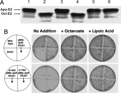
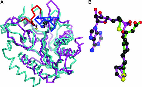
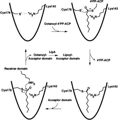
Lipoic acid is essential for the activation of a number of protein complexes involved in key metabolic processes. Growth of Mycobacterium tuberculosis relies on a pathway in which the lipoate attachment group is synthesized from an endogenously produced octanoic acid moiety. In patients with multiple-drug-resistant M. tuberculosis, expression of one gene from this pathway, lipB, encoding for octanoyl-[acyl carrier protein]-protein acyltransferase is considerably up-regulated, thus making it a potential target in the search for novel antiinfectives against tuberculosis. Here we present the crystal structure of the M. tuberculosis LipB protein at atomic resolution, showing an unexpected thioether-linked active-site complex with decanoic acid. We provide evidence that the transferase functions as a cysteine/lysine dyad acyltransferase, in which two invariant residues (Lys-142 and Cys-176) are likely to function as acid/base catalysts. Analysis by MS reveals that the LipB catalytic reaction proceeds by means of an internal thioesteracyl intermediate. Structural comparison of LipB with lipoate protein ligase A indicates that, despite conserved structural and sequence active-site features in the two enzymes, 4'-phosphopantetheine-bound octanoic acid recognition is a specific property of LipB.


Conflict of interest statement

Conflict of interest statement: No conflicts declared.

Figures

Fig. 1.
Fig. 1.
Molecular structure of mtbLipB. (A) Ribbon representation of mtbLipB. Color codes: core β-sheet, yellow; capping β-sheet, orange; α-helices, wrapping the core β-sheet, cyan; other α-helices, brown. The decanoic acid–Cys-176 adduct is shown in ball-and-stick representation, using atom-type colors and bonds in green. (B) Surface presentation of mtbLipB, showing relative electrostatic potentials, using the apbs program incorporated in pymol (see ref. ; www.pymol.org). The visible part of the decanoid acid adduct is shown by sticks in atom-type colors. The orientation has been optimized to provide a view into the mtbLipB active site. Some charged surface residues within the vicinity of the access area into the active site are labeled. (C) mtbLipB active site in ball-and-stick representation, using the color scheme from A. The σA-weighted final 2mFo−DFc electron density covering the decanoic acid moiety is shown at a contour level of 1σ. (D) Schematic presentation of the mtbLipB active site. Specific interactions are limited to Cys-176 and Lys-79. All other interactions are shown schematically; involvement of side chains is in red, and involvement of main chain atoms only is in green.
Fig. 2.
Fig. 2.
Biochemical data. (A) In vitro gel-shift assay. Lane 1, mtbLipB, expressed in E. coli, without octanoy-l-ACP; lane 2, mtbLipB, expressed in M. smegmatis; lane 3, negative control; lane 4, mtbLipB, expressed in E. coli; lane 5, mtbLipB(K142S), expressed in E. coli; lane 6, mtbLipB(C176S), expressed in E. coli. (B) Abilities of wild-type and mutant M. tuberculosis lipB genes to complement growth of an E. coli lipB null mutant strain. The upper row of plates shows the growth behavior of the wild-type E. coli strain JK1 (left top quadrant), the lipB::cat mutant strain ZX221 derived from JK1 (left bottom quadrant), and strain ZX221 carrying the plasmid encoding for the wild-type M. tuberculosis LipB protein (both right quadrants). The lower row of plates similarly shows growth of strain ZX221 carrying plasmid encoding for the mutant M. tuberculosis LipB proteins, either the K142S protein (both left quadrants) or the C176S protein (both right quadrants). Experimental procedures have been described (7).
Fig. 3.
Fig. 3.
Comparison of the structures of LipB and LplA. (A) Superimposed Cα traces of structures of LipB from M. tuberculosis (cyan) and LplA from T. acidophilum (magenta). Enzyme-specific loops are shown in red and blue, respectively. Lys-142 and Cys-176 from LipB and Lys-145 from LplA are shown in ball-and-stick representation. (B) Superposition of decanoid acid (LipB) and lipoyl–AMP (LplA), based on the superimposed protein coordinates from A.
Fig. 4.
Fig. 4.
Structure-based proposal for a LipB reaction mechanism by means of a covalent Cys-176–acyl intermediate. The ε-amino group of the lysine from the lipoyl domain to which octanoate is covalently attached needs to be deprotonated to be catalytically active as a nucleophile. Given that there are neither possible candidate residues, except Lys-142, nor ordered solvent molecules near the active-site center in the LipB structure, we hypothesize there may be proton shuttling between Lys-142 from LipB and the lipoyl domain active lysine (not shown).

References

    1. Perham R. N. Annu. Rev. Biochem. 2000;69:961–1004. - PubMed
    1. Morris T. W., Reed K. E., Cronan J. E., Jr. J. Bacteriol. 1995;177:1–10. - PMC - PubMed
    1. Jordan S. W., Cronan J. E., Jr. Methods Enzymol. 1997;279:176–183. - PubMed
    1. Jordan S. W., Cronan J. E., Jr. J. Bacteriol. 2003;185:1582–1589. - PMC - PubMed
    1. Miller J. R., Busby R. W., Jordan S. W., Cheek J., Henshaw T. F., Ashley G. W., Broderick J. B., Cronan J. E., Jr., Marletta M. A. Biochemistry. 2000;39:15166–15178. - PubMed

Publication types

Associated data

LinkOut - more resources