. 2006 Sep 1;281(35):25062-75.
doi: 10.1074/jbc.M600688200. Epub 2006 May 30.

Cysteine biosynthesis in Trichomonas vaginalis involves cysteine synthase utilizing O-phosphoserine

Affiliations

Cysteine biosynthesis in Trichomonas vaginalis involves cysteine synthase utilizing O-phosphoserine

Gareth D Westrop et al. J Biol Chem. .

Abstract

Trichomonas vaginalis is an early divergent eukaryote with many unusual biochemical features. It is an anaerobic protozoan parasite of humans that is thought to rely heavily on cysteine as a major redox buffer, because it lacks glutathione. We report here that for synthesis of cysteine from sulfide, T. vaginalis relies upon cysteine synthase. The enzyme (TvCS1) can use either O-acetylserine or O-phosphoserine as substrates. The K(m) values of the enzyme for sulfide are very low (0.02 mm), suggesting that the enzyme may be a means of ensuring that sulfide in the parasite is maintained at a low level. T. vaginalis appears to lack serine acetyltransferase, the source of O-acetylserine in many cells, but has a functional 3-phosphoglycerate dehydrogenase and an O-phosphoserine aminotransferase that together result in the production of O-phosphoserine, suggesting that this is the physiological substrate. TvCS1 can also use thiosulfate as substrate. Overall, TvCS1 has substrate specificities similar to those reported for cysteine synthases of Aeropyrum pernix and Escherichia coli, and this is reflected by sequence similarities around the active site. We suggest that these enzymes are classified together as type B cysteine synthases, and we hypothesize that the use of O-phosphoserine is a common characteristic of these cysteine synthases. The level of cysteine synthase in T. vaginalis is regulated according to need, such that parasites growing in an environment rich in cysteine have low activity, whereas exposure to propargylglycine results in elevated cysteine synthase activity. Humans lack cysteine synthase; therefore, this parasite enzyme could be an exploitable drug target.


Figures
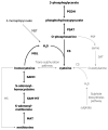
Fig. 1
Fig. 1
Cysteine biosynthesis in T. vaginalis. Enzymes present in T. vaginalis and reactions catalysed by them (this study and previous published work) are shown in bold, enzymes predicted to be present are given in normal type. Enzymes apparently absent from T. vaginalis are in grey type. See Table 1 for key to abbreviations. MGL, methionine γ-lyase; SAM-MT, S-adenosylmethione-dependent methyltransferase (49); SAHH, S-adenosylhomocysteine hydrolase (50).
Fig. 2
Fig. 2
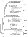
Phylogenetic tree of CS sequences. Sequences were aligned with AlignX (Vector NTI 9.0, Invitrogen) and all regions with gaps and inserts/extensions were removed. Phylogenetic analyses were conducted using MEGA version 3.0 (24) on 207 conserved positions. A bootstrap test was performed with the Neighbour-joining method (500 replicates, seed 74307). Bootstrap values for interior branches of < 95% are shown. Protein accession numbers are shown in brackets. The bar shows the number of substitutions per amino acid position.
Fig. 3
Fig. 3
Multiple alignments of protein sequences of various cysteine synthases. Sequences were aligned with AlignX (Vector NTI 9.0, Invitrogen) and edited by reference to published alignments based on 3D structures of S. typhimurium CS type A (StCS-A) (18), E. coli CS type B (EcCS-B) (20), A. pernix CS (22) and A. thaliana CS (32). Blocks of conserved residues are indicated by white on a black background and blocks of similar residues by white on a grey background. Sequences are arranged into 6 groups: 1, bacterial CS type A sequences; 2, protozoan CS, type A-related; 3, plant CSs; 4, bacterial CS type B sequences; 5, CS, type B-related; 6, archaeon CSs. EcCS-A, E. coli CS type A (NP-311319); StCS-A, S. typhimurium CS type A (NP_461365]); EhCS, E. histolytica CS (BAA21916); LmCS, L. major CS (LmjF36.3590); AtCS, A. thaliana CS (P47998); AtCS chlor., A. thaliana chloroplast CS (P47999); AtCS mito., A. thaliana mitochondrial CS (Q43725); EcCS-B., E. coli CS type B (NP_311319); StCS-B, S. typhimurium CS type B (NP_461375); GsCS-B, G. sulfurreducens CS type B (AAR36549); TvCS1, T. vaginalis CS ([95238.m00105); ApCS, A. pernix CS (NP_148041); PfCS, P. furiosus CS (CAB49296). (a), Conserved N-terminal amino acid residues. Active site lysine is indicated by symbol ▲. (b), loop region amino acid residues (located between β8 and β9). Structural features identified in plant and bacterial type A CS proteins are indicated by symbols above the top sequence: *, highly conserved sequence; ‡,residues implicated (by mutagenesis of AtCS) in binding of serine acetyltransferase to plant CS proteins. Structural features identified in bacterial type B and thermophile CS proteins are indicated by symbols below the bottom sequence: •, loop forming part of active site pocket (20,22); , conserved positively charged residue implicated (by mutagenesis) in binding of O-phosphoserine to thermophile CS proteins (22).
Fig. 3
Fig. 3
Multiple alignments of protein sequences of various cysteine synthases. Sequences were aligned with AlignX (Vector NTI 9.0, Invitrogen) and edited by reference to published alignments based on 3D structures of S. typhimurium CS type A (StCS-A) (18), E. coli CS type B (EcCS-B) (20), A. pernix CS (22) and A. thaliana CS (32). Blocks of conserved residues are indicated by white on a black background and blocks of similar residues by white on a grey background. Sequences are arranged into 6 groups: 1, bacterial CS type A sequences; 2, protozoan CS, type A-related; 3, plant CSs; 4, bacterial CS type B sequences; 5, CS, type B-related; 6, archaeon CSs. EcCS-A, E. coli CS type A (NP-311319); StCS-A, S. typhimurium CS type A (NP_461365]); EhCS, E. histolytica CS (BAA21916); LmCS, L. major CS (LmjF36.3590); AtCS, A. thaliana CS (P47998); AtCS chlor., A. thaliana chloroplast CS (P47999); AtCS mito., A. thaliana mitochondrial CS (Q43725); EcCS-B., E. coli CS type B (NP_311319); StCS-B, S. typhimurium CS type B (NP_461375); GsCS-B, G. sulfurreducens CS type B (AAR36549); TvCS1, T. vaginalis CS ([95238.m00105); ApCS, A. pernix CS (NP_148041); PfCS, P. furiosus CS (CAB49296). (a), Conserved N-terminal amino acid residues. Active site lysine is indicated by symbol ▲. (b), loop region amino acid residues (located between β8 and β9). Structural features identified in plant and bacterial type A CS proteins are indicated by symbols above the top sequence: *, highly conserved sequence; ‡,residues implicated (by mutagenesis of AtCS) in binding of serine acetyltransferase to plant CS proteins. Structural features identified in bacterial type B and thermophile CS proteins are indicated by symbols below the bottom sequence: •, loop forming part of active site pocket (20,22); , conserved positively charged residue implicated (by mutagenesis) in binding of O-phosphoserine to thermophile CS proteins (22).
Fig. 4
Fig. 4
Recombinant proteins used in activity analyses. Lane 1, rTvCS1 (34.7 kDa); lane 2, rTvPSAT11 (44.1 kDa); lane 3, rTvPGDH11 (45.1 kDa). All ~5 μg/lane.
Fig. 5
Fig. 5
Variation of cysteine synthase activity of rTvCS1 with substrate concentration. Inset: double reciprocal plot. a, 1-100 mM O-acetylserine with 3 mM sodium sulphide (Na2S); b, 0.1–10 mM sodium sulphide with 100 mM O-acetylserine; c, 50–270 mM O-phosphoserine with 1 mM sodium sulphide; d, 0.01–1 mM sodium sulphide with 80 mM O-phosphoserine.
Fig. 6
Fig. 6
Changes in CS protein, enzymic activity and mRNA levels in T. vaginalis grown under different conditions. (a) The protein expression levels determined by western blotting: TvCS, cysteine synthase; TvTR, thioredoxin reductase (14). (b) CS activities, means ± SD from 3 experiments in μmol/min/mg protein, determined at 37°C in 0.2 ml reactions containing 15 mM O-acetylserine, 3 mM sodium sulphide and 10 μg soluble protein from T. vaginalis. (c) The levels of mRNA assessed by quantitation of 32P hybridization using a phosphoimager and shown relative to the levels in T. vaginalis grown under standard conditions. The variation in growth conditions were: lane 1, control; lane 2, without ascorbate, lane 3; with added 10 mM cysteine; lane 4, with 5 μM propargylglycine. Northern blot analysis was not carried out for the propargylglycine-treated cells.

References

    1. Petrin D, Delgaty K, Bhatt R, Garber G. Clinical. Microbiol. Rev. 1998;11:300–317. - PMC - PubMed
    1. World Health Organisation Global prevalence and incidence of selected curable sexually transmitted diseases: overview and estimates. 2001 WHO/CDS/CSR/EDC/2001.10.
    1. Sorvillo F, Smith L, Kerndt P, Ash L. Emerg. Infect. Dis. 2001;7:927–932. - PMC - PubMed
    1. Baldauf SL, Roger AJ, Wenk-Siefert I, Doolittle WF. Science. 2000;290:972–977. - PubMed
    1. Horner DS, Embley TM. Mol. Biol. Evol. 2001;18:1970–1975. - PubMed

Publication types

LinkOut - more resources