. 2006 Jun 5;173(5):719-31.
doi: 10.1083/jcb.200511125. Epub 2006 May 30.

An intimate collaboration between peroxisomes and lipid bodies

Affiliations

An intimate collaboration between peroxisomes and lipid bodies

Derk Binns et al. J Cell Biol. .

Abstract

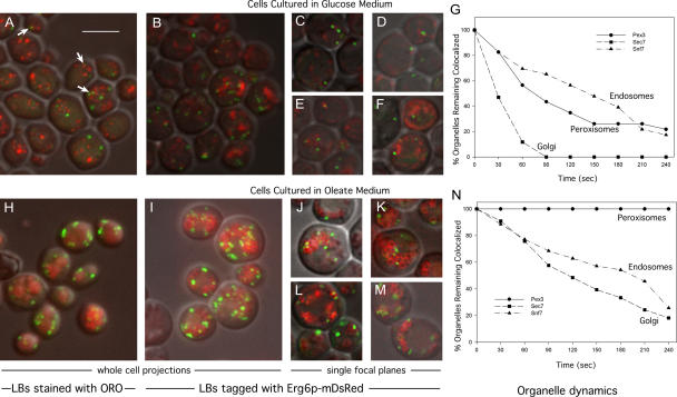
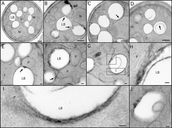
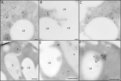
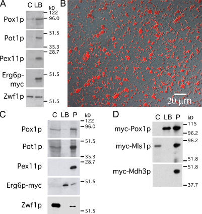
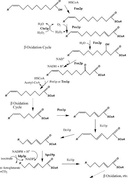
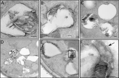
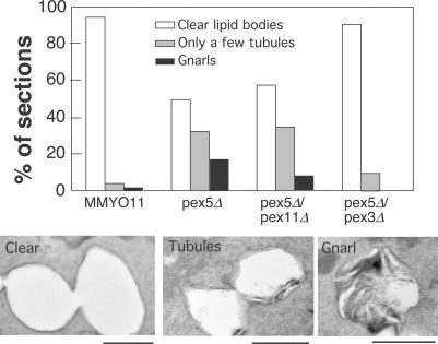
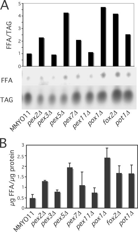
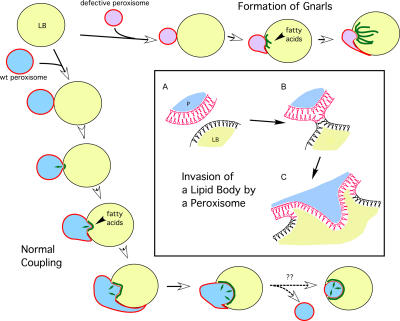
Although peroxisomes oxidize lipids, the metabolism of lipid bodies and peroxisomes is thought to be largely uncoupled from one another. In this study, using oleic acid-cultured Saccharomyces cerevisiae as a model system, we provide evidence that lipid bodies and peroxisomes have a close physiological relationship. Peroxisomes adhere stably to lipid bodies, and they can even extend processes into lipid body cores. Biochemical experiments and proteomic analysis of the purified lipid bodies suggest that these processes are limited to enzymes of fatty acid beta oxidation. Peroxisomes that are unable to oxidize fatty acids promote novel structures within lipid bodies ("gnarls"), which may be organized arrays of accumulated free fatty acids. However, gnarls are suppressed, and fatty acids are not accumulated in the absence of peroxisomal membranes. Our results suggest that the extensive physical contact between peroxisomes and lipid bodies promotes the coupling of lipolysis within lipid bodies with peroxisomal fatty acid oxidation.


Figures

Figure 1.
Figure 1.
Peroxisomes decorate lipid bodies. MMYO11α expressing Pot1p (thiolase)-GFP (A–F and H–M) or other GFP-tagged proteins (G and N) was grown in SD medium (2% glucose; A–G) or oleate medium (H–N). Cells were either fixed and stained with oil red O (ORO; A and H) or expressed Erg6p-mDsRed (B–G and I–N). A, B, H, and I represent projections through entire cells, whereas C–F and J–M are images of single planes. Lipid bodies (LBs) are larger and more abundant in oleate medium (J–M). Examples of peroxisomes associating with lipid bodies are indicated by arrows in A. For G and N, Erg6p-mDsRed was introduced into strains expressing GFP-tagged Pex3p, Sec7p, or Snf7p as indicated. Individual GFP-tagged organelles (20–30 per strain) that were associated with lipid bodies in glucose (G) or oleate (N) cultures were observed continually over 4 min and scored for release from lipid bodies. For clarity, the graphs show loss of organelles at 30-s intervals. Bar, 5 μm (applies to all images).
Figure 2.
Figure 2.
Ultrastructure of lipid bodies and contacts with peroxisomes. Cells were grown for 18–21 h in oleate medium and processed for EM. (A–D) Aspects of lipid body structure are illustrated, particularly their connections to each other (A and B) and inclusions (C and D). Arrows in B illustrate valvelike structures; arrows in C and D illustrate inclusions. (E–H) Lipid bodies in contact with peroxisomes are shown, illustrating electron-dense material at the site of contact (arrows). Note a peroxisome in contact with three lipid bodies (E). A lipid body making contact with two peroxisomes, one of which has extended a tail around it, and the other has breached the periphery and is extending a process (pexopodium) into the core (G). The boxed areas are shown in higher resolution in H and I. Another putative pexopodium is shown in J. LB, lipid body; M, mitochondria; N, nucleus, P, peroxisome. Bars (A–D), 500 nm; (E–J), 50 nm.
Figure 3.
Figure 3.
Lipid body inclusions contain Pox1p. MMYO11α was grown overnight in oleate medium and processed for immunogold EM. (A–C) Anti-Pot1p antibody illustrates peroxisomes making extensive contacts with the lipid body. (D–F) Anti-Pox1p antibodies stain peroxisomes (D) but also stain inclusions (pexopodia) in lipid bodies. We have not detected the immunoreactivity of pexopodia with anti-Pot1p. LB, lipid body; P, peroxisome; V, vacuole. Bars, 100 μm.
Figure 4.
Figure 4.
Purified lipid bodies contain Pox1p. (A) A PNS from cells expressing Erg6p-myc was grown in oleic acid and gently centrifuged to float lipid bodies. The lipid body fraction and underlying supernatant were analyzed for organelle markers. Peroxisomal proteins but not cytosolic Zwf1p were enriched in the lipid body fraction. (B) Lipid bodies were purified and stained with oil red O. (C) Purified lipid bodies and an organellar pellet were analyzed for peroxisome markers. An equal percentage of cytosol, washed lipid bodies, and pellet (C, LB, and P, respectively) were analyzed by immunoblotting. Pox1p but not Pot1p or Pex11p is visible in the lipid body fraction. (D) As in C, except cells expressed myc-tagged Pox1p, Mls1p, or Mdh3p as indicated, and they were cultured and processed in parallel.
Figure 5.
Figure 5.
Peroxisomal β oxidation of oleic acid in S. cerevisiae. The first round is shown in detail. The enzymes in bold are the only peroxisomal proteins identified in the lipid body proteome. These may represent the major proteins of pexopodia.
Figure 6.
Figure 6.
Gnarls. (A–F) Gnarled lipid bodies from Δpex5 are illustrated. A higher resolution image from the boxed area in E is shown in F. Arrow points to the pattern of electron-dense planes, 7 μm apart, which may be sheets of fatty acid–enriched membranes. Bars (A–C and F), 100 nm; (D and E), 500 nm.
Figure 7.
Figure 7.
Pex3p is essential to promote gnarls. 50 cells of the indicated strains (blinded) were scored for lipid bodies that were clear, contained at most a few tubules, or contained gnarls. Examples of each are shown below. Bars, 500 nm.
Figure 8.
Figure 8.
Peroxisomal mutants cause fatty acid accumulation in lipid bodies. Lipid bodies were purified from the indicated strains and subjected to thin layer chromatography. (A) Spots were visualized and quantified after charring, and the ratio of free fatty acids to triacylglycerols are plotted. (B) Free fatty acid amounts were quantified by densitometric scanning relative to free oleic acid standards and were normalized to total lipid body protein. Results shown were based on three independent experiments. Means and SEM (error bars) are plotted.
Figure 9.
Figure 9.
Scheme depicting the development and dynamics of peroxisome–lipid body interactions. (left and bottom) Peroxisomes bind to lipid bodies (LBs) and stimulate the breakdown of triglycerides, releasing fatty acids (green lines) that get imported and oxidized by peroxisomes. Peroxisome invasion into the lipid body is also shown. (top) Peroxisomes that are unable to perform β oxidation cause the accumulation of fatty acids, resulting in gnarls. (box) Peroxisome invasion into lipid bodies. (A) A peroxisome (P) with its phospholipid bilayer approaches the lipid body monolayer. (B) A hemifusion event occurs, resulting in invasion of the lipid core by the peroxisomal membrane inner leaflet (C). Small arrows indicate the flux of fatty acids into peroxisomes for oxidation.

References

    1. Athenstaedt, K., and G. Daum. 2005. Tgl4p and Tgl5p, two triacylglycerol lipases of the yeast Saccharomyces cerevisiae are localized to lipid particles. J. Biol. Chem. 280:37301–37309. - PubMed
    1. Athenstaedt, K., D. Zweytick, A. Jandrositz, S.D. Kohlwein, and G. Daum. 1999. Identification and characterization of major lipid particle proteins of the yeast Saccharomyces cerevisiae. J. Bacteriol. 181:6441–6448. - PMC - PubMed
    1. Baerends, R.J., S.W. Rasmussen, R.E. Hilbrands, M. van der Heide, K.N. Faber, P.T. Reuvekamp, J.A. Kiel, J.M. Cregg, I.J. van der Klei, and M. Veenhuis. 1996. The Hansenula polymorpha PER9 gene encodes a peroxisomal membrane protein essential for peroxisome assembly and integrity. J. Biol. Chem. 271:8887–8894. - PubMed
    1. Bascom, R.A., H. Chan, and R.A. Rachubinski. 2003. Peroxisome biogenesis occurs in an unsynchronized manner in close association with the endoplasmic reticulum in temperature-sensitive Yarrowia lipolytica Pex3p mutants. Mol. Biol. Cell. 14:939–957. - PMC - PubMed
    1. Battaile, K.P., J. Molin-Case, R. Paschke, M. Wang, D. Bennett, J. Vockley, and J.J. Kim. 2002. Crystal structure of rat short chain acyl-CoA dehydrogenase complexed with acetoacetyl-CoA: comparison with other acyl-CoA dehydrogenases. J. Biol. Chem. 277:12200–12207. - PubMed

Publication types

MeSH terms