. 2006 Jun 5;173(5):695-707.
doi: 10.1083/jcb.200601011. Epub 2006 May 30.

Asi1 is an inner nuclear membrane protein that restricts promoter access of two latent transcription factors

Affiliations

Asi1 is an inner nuclear membrane protein that restricts promoter access of two latent transcription factors

Mirta Boban et al. J Cell Biol. .

Abstract

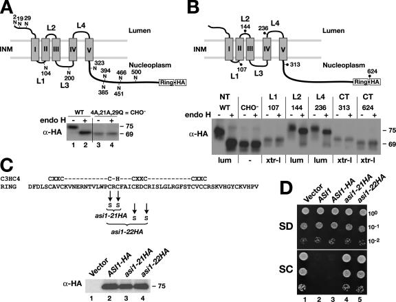
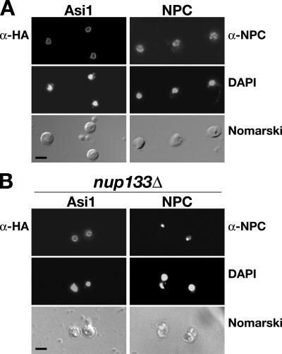
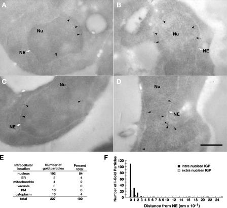
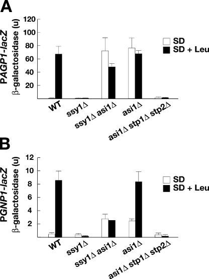
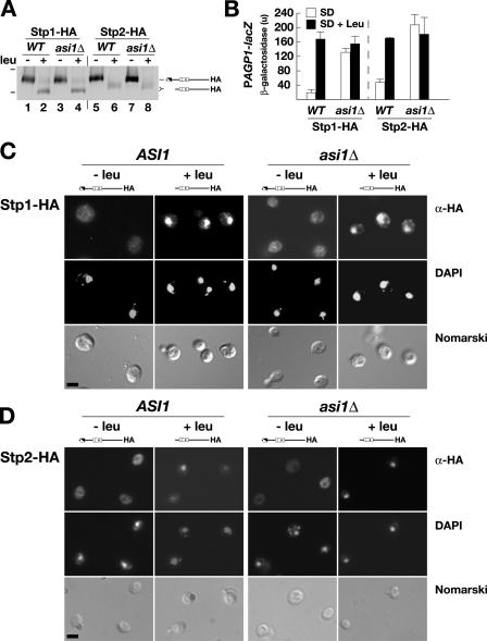
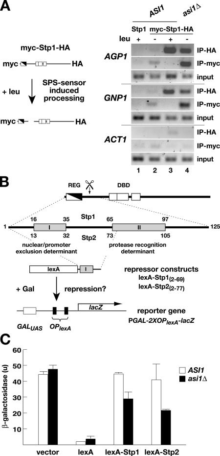
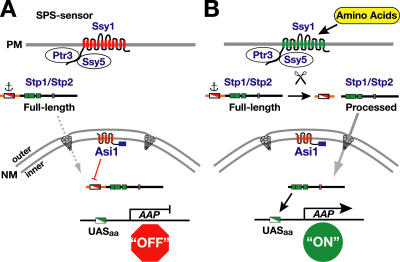
Stp1 and Stp2 are homologous transcription factors in yeast that are synthesized as latent cytoplasmic precursors with NH2-terminal regulatory domains. In response to extracellular amino acids, the plasma membrane-localized Ssy1-Ptr3-Ssy5 (SPS) sensor endoproteolytically processes Stp1 and Stp2, an event that releases the regulatory domains. The processed forms of Stp1 and Stp2 efficiently target to the nucleus and bind promoters of amino acid permease genes. In this study, we report that Asi1 is an integral component of the inner nuclear membrane that maintains the latent characteristics of unprocessed Stp1 and Stp2. In cells lacking Asi1, full-length forms of Stp1 and Stp2 constitutively induce SPS sensor-regulated genes. The regulatory domains of Stp1 and Stp2 contain a conserved motif that confers Asi1-mediated control when fused to an unrelated DNA-binding protein. Our results indicate that latent precursor forms of Stp1 and Stp2 inefficiently enter the nucleus; however, once there, Asi1 restricts them from binding SPS sensor-regulated promoters. These findings reveal an unanticipated role of inner nuclear membrane proteins in controlling gene expression.


Figures

Figure 1.
Figure 1.
Structural and functional analysis of Asi1. (A) Asi1 is a glycoprotein. Schematic presentation of the predicted membrane topology of Asi1; the 11 possible NH2-linked glycosylation sites are shown. The gray boxes depict the five hydrophobic domains (I–V), and the COOH-terminal RING domain and HA epitope tag are indicated. Strain YMH349 (asi1Δ ssy1Δ leu2) carrying either plasmid pAZ002 (ASI1-HA) or pAZ020 (ASI1-HA-CHO ) was grown in SC. Extracts of total cell protein were prepared, treated with endoH as indicated, and resolved by SDS-PAGE and immunoblotted with monoclonal anti-HA antibody. The numbers to the right of immunoblots are the molecular mass (in kilodaltons) of protein bands calculated based on the migration of protein standards. INM, inner nuclear membrane. (B) Membrane topology of Asi1. Schematic representation of Asi1. Positions of the suc2 topological reporter cassette insertions are indicated by dots; the numbers refer to the amino acids to which the insertions were fused. Native Asi1-HA (pAZ014), glycosylation minus Asi1-HA-CHO (pAZ020), and suc2 cassette gene fusion proteins Asi1-107 (pAZ034), Asi1-144 (pAZ035), Asi1-236 (pAZ037), Asi1-313 (pAZ039), and Asi1-624 (pAZ073) were expressed in YMH349. Cells were grown in SD, and extracts were prepared and analyzed as in A. The experimentally determined topological orientation of each reporter is indicated (lum, lumenal; xtr-l, extralumenal). NT, NH2 terminus; CT, COOH terminus. (C and D) The C3HC4-like RING motif (amino acids 568–608) is required for function. WT, wild type. (C) Alleles asi1-21HA (pAZ015) and asi1-22HA (pAZ016) encode mutant proteins with cysteine 583 and 585 and cysteine 583, 585, 589, and 592 replaced by serine, respectively. Immunoblot analysis of extracts from strain YMH349 carrying plasmids pRS202 (vector), pAZ014 (ASI1-HA), pAZ015 (asi1-21HA), or pAZ016 (asi1-22HA) grown in SD. Proteins were resolved on 10% SDS-PAGE gels and blotted with anti-HA antibody. (D) 10-fold dilution series of cell suspensions of strains as in C and YMH349 carrying pMH4 (ASI1) were spotted onto plates containing SD or SC, incubated for 3 d at 30°C, and photographed.
Figure 2.
Figure 2.
Asi1 is a component of the nuclear envelope. Indirect immunolocalization of Asi1 (left) and nuclear pore complexes (NPCs; right) was performed with anti-HA (α-HA) and anti-NPC (α-NPC) monoclonal antibodies, respectively. (A) Strain PLY1314 (asi1Δ) carrying pAZ014 (ASI1-HA) was grown on SC (−ura). The pattern of NPC staining was examined in strain AZY262 (ASI1) grown in YPD. (B) Asi1 is not directly associated with NPCs. Strain nup133 (nup133Δ) carrying pAZ014 (ASI1-HA) was grown on SC. (top to bottom) Monoclonal α-HA (12CA5 or 3F10) or α-NPC (anti-rNup153) antibody-dependent AlexaFluor488 fluorescence; DAPI staining; cells viewed by Nomarski optics. Bars, 5 μm.
Figure 3.
Figure 3.
Asi1 is a component of the inner nuclear membrane. Strain PLY1314 (asi1Δ) carrying pAZ014 (ASI1-HA) was grown in SC (−ura). Cells were fixed and prepared for analysis as described in Materials and methods. (A–D) Immunoelectron micrographs of yeast cell nuclei (Nu). The white arrows indicate the nuclear envelope (NE), and the black arrowheads show the locations of Asi1-HA–dependent labeling of gold particles. Quantification of immunogold labeling. Bar, 0.5 μm. (E) Intracellular distribution of immunogold particles in 44 cells decorated with at least two gold particles. PM, plasma membrane. (F) The distance of 165 immunogold particles (IGPs) from the nuclear membrane.
Figure 4.
Figure 4.
Null alleles of ASI1 derepress SPS sensor–regulated genes in a strictly Stp1- and Stp2-dependent manner. The expression of two SPS sensor–dependent genes, AGP1 (A) and GNP1 (B), was monitored by measuring β-galactosidase activity (in units) of PAGP1-lacZ and PGNP1-lacZ reporter constructs. Wild-type (WT; CAY29), ssy1Δ (CAY91), ssy1Δ asi1Δ (CAY206), asi1Δ (PLY1313), and asi1Δ stp1Δ stp2Δ (CAY152) strains carrying either plasmid YCpAGP1-lacZ (PAGP1-lacZ) or pCA227 (PGNP1-lacZ) were grown in SD or SD supplemented with leucine (SD + Leu). β-galactosidase activity present in three independent transformants of each strain was quantified; error bars indicate one standard deviation.
Figure 5.
Figure 5.
Characterization of the SPS sensor signaling pathway in asi1-null mutant strains. (A) Stp1 and Stp2 processing occurs independently of Asi1. ASI1 (PLY127) and asi1Δ (PLY1327) strains were transformed with pCA030 (PAGP1-lacZ) and pCA047 (Stp1-HA) or pCA111 (Stp2-HA). Cells were grown in SD medium (−leu), and, where indicated, leucine was added 30 min before harvest (+leu). Extracts were resolved by SDS-PAGE and immunoblotted with anti-HA. The immunoreactive forms of Stp1-HA and Stp2-HA present in the cell extracts are schematically represented (right), and the 83- and 62-kD protein standards (bars, left) are indicated at their corresponding positions of migration. WT, wild type. (B) The levels of β-galactosidase activity (in units) resulting from expression of the PAGP1-promoted β-galactosidase gene were assessed in strains as in A. The activity in three independent transformants was quantified; error bars indicate one standard deviation. (C and D) Indirect immunolocalization of Stp1-HA (C) and Stp2-HA (D) in ASI1 and asi1Δ cells was performed with anti-HA monoclonal antibodies. Cells were grown in SD to an OD600 of 0.7, the cultures were split into two equal aliquots, and leucine was added to one. The cultures were incubated by shaking at 30°C for an additional 30 min, and the cells were fixed. (top to bottom) α-HA monoclonal antibody-dependent AlexaFluor488 fluorescence; DAPI staining; and cells viewed by Nomarski optics. Strains CAY28 (ASI1) and PLY1314 (asi1Δ) carrying pCA078 (STP1-HA; C) and strains CAY29 (ASI1) and PLY1313 (asi1Δ) carrying pMB30 (STP2-HA; D) were used. The full-length (uninduced; −leu) and processed (induced; +leu) forms of Stp1-HA and Stp2-HA present in cells are schematically depicted above the appropriate panels. Bars, 5 μm.
Figure 6.
Figure 6.
Unprocessed Stp1 and Stp2 are excluded from SPS sensor–regulated promoters in an Asi1-dependent manner. (A) ChIP analysis of Stp1 association with AGP1 and GNP1 promoters before and after amino acid–induced processing. Schematic diagram of the double epitope-tagged myc-Stp1-HA protein and SPS sensor–dependent processing. Cultures of ASI1 (CAY60) and asi1Δ (CAY150) carrying plasmid pMB10 (myc-Stp1-HA) were grown in SD medium (−leu), and, where indicated, leucine was added 30 min before harvest (+leu). Cell lysates were prepared and split into two equal portions, and each portion was analyzed by ChIP using anti-myc or anti-HA antibody. The ability to amplify the ACT1 promoter was monitored to control nonspecific immunoprecipitation. The size of amplified fragments are as follows: AGP1, 246 bp; GNP1, 313 bp; and ACT1, 274 bp. (B) Diagram of Stp1 and Stp2 and the repression assay used to assess Asi1-dependent promoter access. Stp1 and Stp2 contain two distinct regions (I and II) of sequence conservation within their NH2-terminal regulatory domains (REG) and DNA-binding domains (DBD). Regions I and II are indicated in the enlargement of the NH2-terminal domain (amino acids 1–125). Region I is required to prevent unprocessed full-length forms from efficiently entering the nucleus and binding promoters. Region II is required for amino acid–induced endoproteolytic processing (scissors). Gene fusion constructs directing the expression of the DNA-binding protein lexA fused to NH2-terminal Stp1(2–69) (pMB16) and Stp2(2–77) (pMB17) Region I–containing sequences are shown. In the absence of lexA, the reporter gene PGAL1-2XOPlexA-lacZ (pMB18) is fully inducible by galactose; if lexA is present and able to localize to the nucleus, the induction is reduced. (C) The ability of lexA fusion proteins to enter the nucleus and bind to lexA operators (OPlexA) was assessed by measuring the levels of β-galactosidase activity (in units) in ASI1 (PLY127) and asi1Δ (PLY1327) strains carrying pMB18 (PGAL1-2XOPlexA-lacZ) and pCA160 (vector), pCA153 (lexA), pMB16 (lexA-Stp1), or pMB17 (lexA-Stp2). Strains were grown in SC and induced with galactose. The activity in three independent transformants was quantified; error bars indicate one standard deviation.
Figure 7.
Figure 7.
Model of the SPS-sensing pathway and the role of Asi1 in maintaining the latency of Stp1 and Stp2. (A) In the absence of inducing amino acids, the SPS sensor (Ssy1–Ptr3–Ssy5) of extracellular amino acids is present in the plasma membrane (PM) in its preactivation conformation (red). The transcription factors Stp1 and Stp2 are synthesized as inactive precursors that localize to the cytosol as a result of the presence of a cytoplasmic retention signal (anchor) that prevents the unprocessed full-length forms to efficiently enter the nucleus. In the absence of Asi1, full-length Stp1 and Stp2 enter the nucleus (dashed arrow) at rates sufficient to derepress amino acid permease (AAP) gene expression. The ability of Asi1 located in the inner nuclear membrane (NM) to prevent transcription is dependent on the nucleoplasmically oriented COOH-terminal RING motif (blue box) and on the presence of the sequences of Stp1 and Stp2 (Region I) in the NH2-terminal regulatory domain (red and white box). Consequently, there are low levels of amino acid permeases in the plasma membrane. (B) In the presence of inducing amino acids, the SPS sensor is activated (green), leading to the endoproteolytic processing of Stp1 and Stp2 (scissors). The shorter activated forms of Stp1 and Stp2 are efficiently targeted to the nucleus (solid arrow), where they bind SPS sensor–regulated promoters (DNA-binding domains; green boxes) and induce their transcription. The increased transcription of amino acid permease genes results in a concomitant increase in amino acid permeases in the plasma membrane, and cells exhibit induced rates of amino uptake.

References

    1. Abdel-Sater, F., M. El Bakkoury, A. Urrestarazu, S. Vissers, and B. André. 2004. Amino acid signaling in yeast: casein kinase I and the Ssy5 endoprotease are key determinants of endoproteolytic activation of the membrane-bound Stp1 transcription factor. Mol. Cell. Biol. 24:9771–9785. - PMC - PubMed
    1. Andréasson, C., and P.O. Ljungdahl. 2002. Receptor-mediated endoproteolytic activation of two transcription factors in yeast. Genes Dev. 16:3158–3172. - PMC - PubMed
    1. Andréasson, C., and P.O. Ljungdahl. 2004. The N-terminal regulatory domain of Stp1p is modular and fused to an artificial transcription factor confers full SPS-sensor control. Mol. Cell. Biol. 24:7503–7513. - PMC - PubMed
    1. Andréasson, C., S. Heessen, and P.O. Ljungdahl. 2006. Regulation of transcription factor latency by receptor-activated proteolysis. Genes Dev. In press. - PMC - PubMed
    1. Antebi, A., and G.R. Fink. 1992. The yeast Ca(2+)-ATPase homologue, PMR1, is required for normal Golgi function and localizes in a novel Golgi-like distribution. Mol. Biol. Cell. 3:633–654. - PMC - PubMed

Publication types

MeSH terms