

Central projections of melanopsin-expressing retinal ganglion cells in the mouse
- PMID: 16736474
- PMCID: PMC2885916
- DOI: 10.1002/cne.20970
Central projections of melanopsin-expressing retinal ganglion cells in the mouse
Abstract
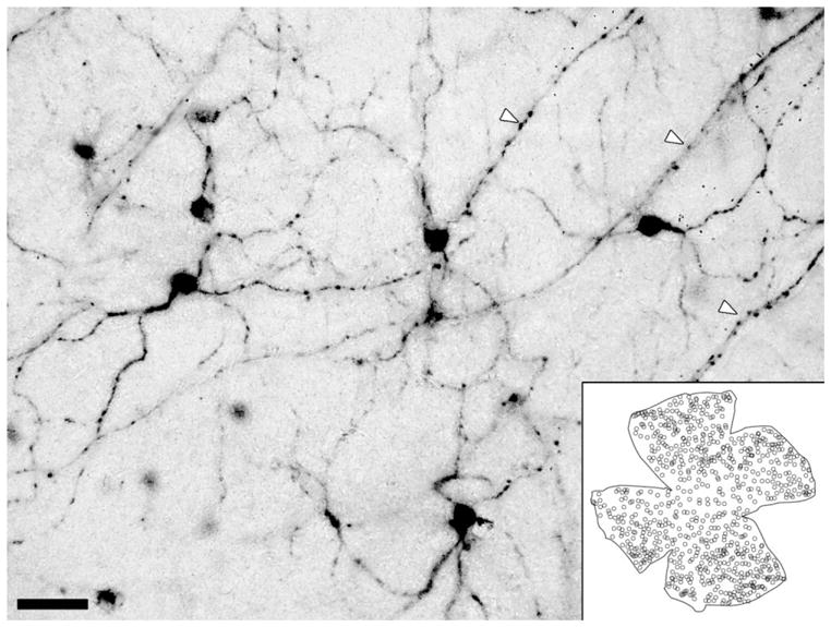
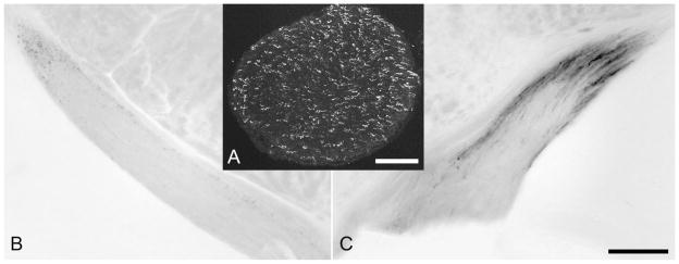
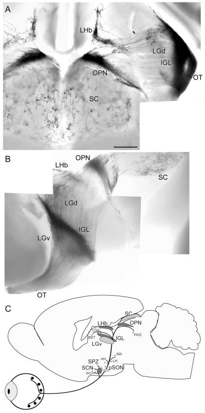
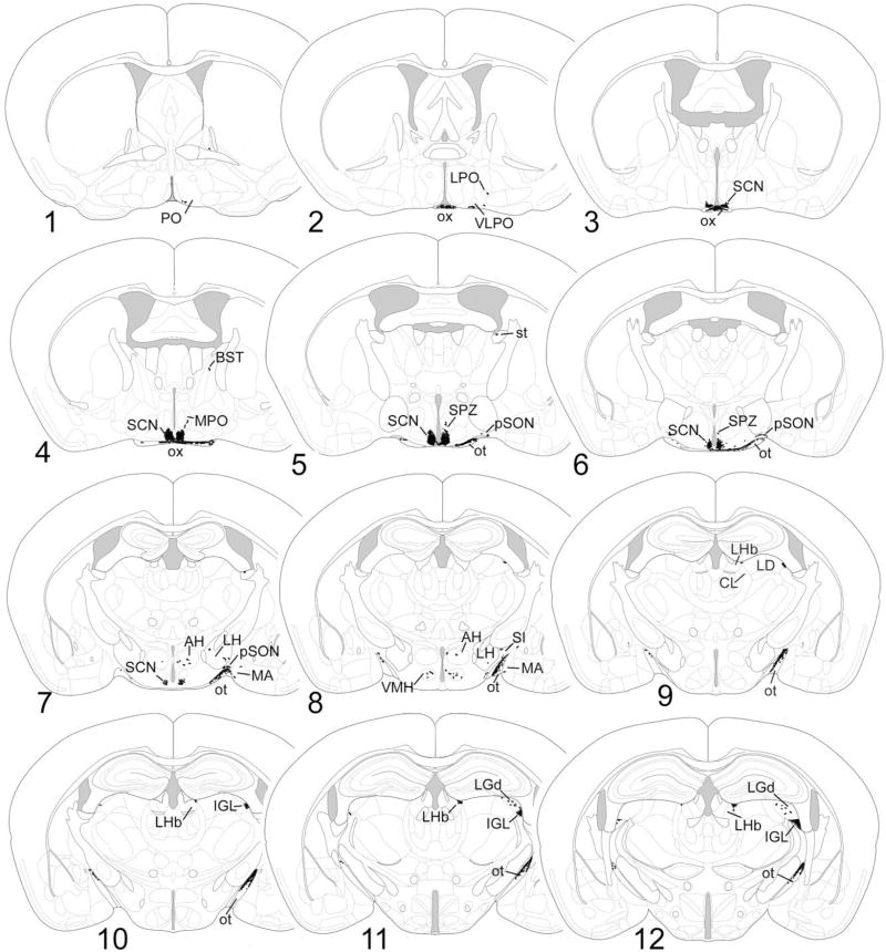
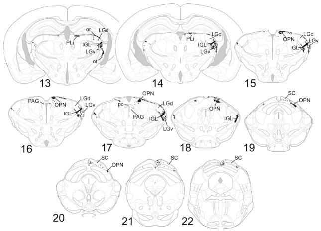
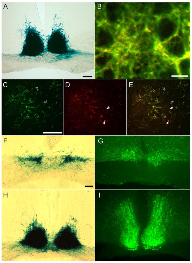
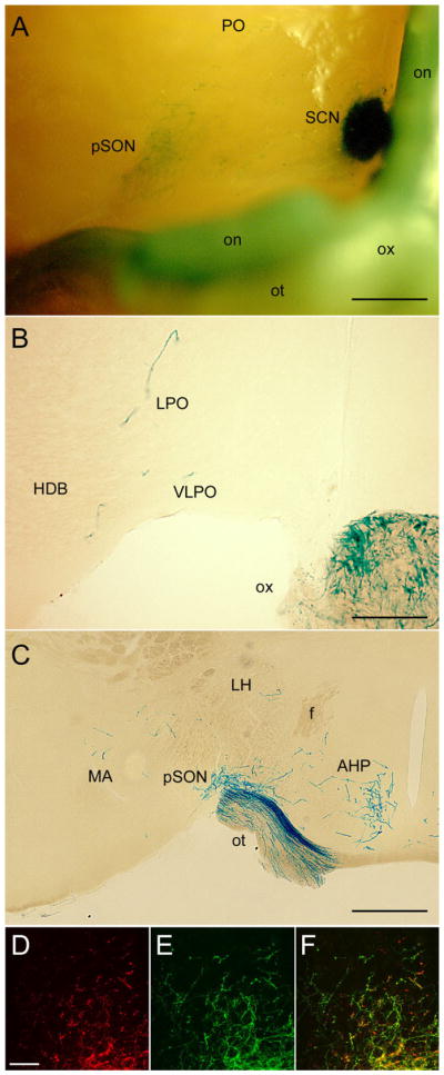
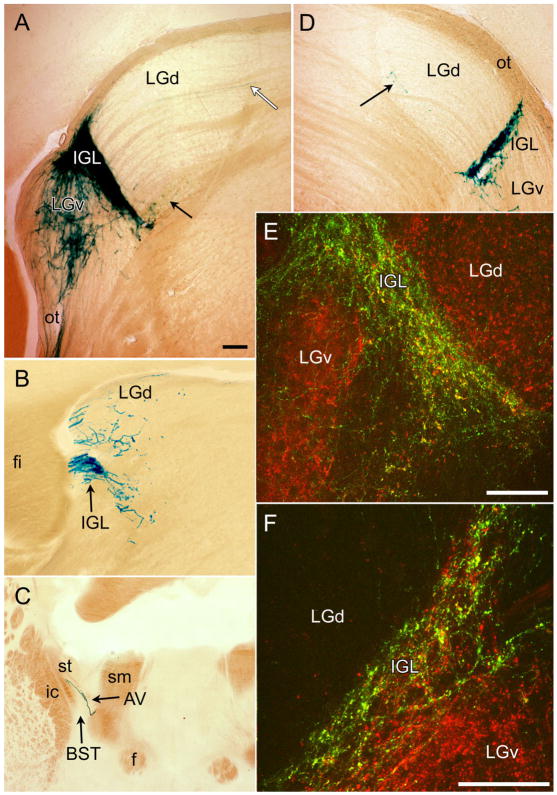
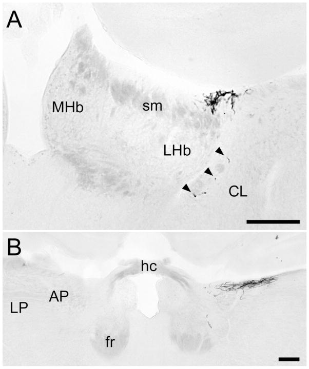
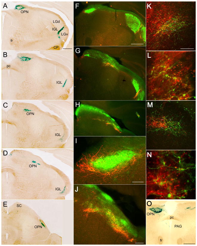
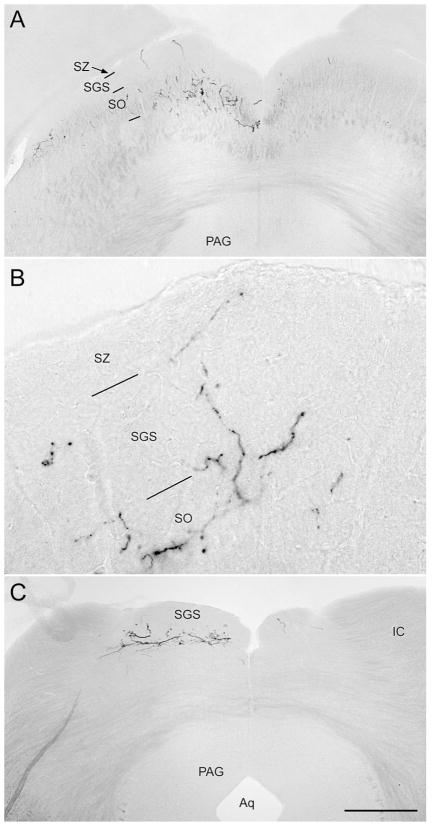
A rare type of ganglion cell in mammalian retina is directly photosensitive. These novel retinal photoreceptors express the photopigment melanopsin. They send axons directly to the suprachiasmatic nucleus (SCN), intergeniculate leaflet (IGL), and olivary pretectal nucleus (OPN), thereby contributing to photic synchronization of circadian rhythms and the pupillary light reflex. Here, we sought to characterize more fully the projections of these cells to the brain. By targeting tau-lacZ to the melanopsin gene locus in mice, ganglion cells that would normally express melanopsin were induced to express, instead, the marker enzyme beta-galactosidase. Their axons were visualized by X-gal histochemistry or anti-beta-galactosidase immunofluorescence. Established targets were confirmed, including the SCN, IGL, OPN, ventral division of the lateral geniculate nucleus (LGv), and preoptic area, but the overall projections were more widespread than previously recognized. Targets included the lateral nucleus, peri-supraoptic nucleus, and subparaventricular zone of the hypothalamus, medial amygdala, margin of the lateral habenula, posterior limitans nucleus, superior colliculus, and periaqueductal gray. There were also weak projections to the margins of the dorsal lateral geniculate nucleus. Co-staining with the cholera toxin B subunit to label all retinal afferents showed that melanopsin ganglion cells provide most of the retinal input to the SCN, IGL, and lateral habenula and much of that to the OPN, but that other ganglion cells do contribute at least some retinal input to these targets. Staining patterns after monocular enucleation revealed that the projections of these cells are overwhelmingly crossed except for the projection to the SCN, which is bilaterally symmetrical.
(c) 2006 Wiley-Liss, Inc.
Figures
References
-
- Abrahamson EE, Moore RY. Suprachiasmatic nucleus in the mouse: retinal innervation, intrinsic organization and efferent projections. Brain Res. 2001;916:172–191. - PubMed
-
- Alvarez-Lopez C, Cernuda-Cernuda R, Paniagua MA, Alvarez-Viejo M, Fernandez-Lopez A, Garcia-Fernandez JM. The transcription factor CREB is phosphorylated in neurons of the piriform cortex of blind mice in response to illumination of the retina. Neurosci Lett. 2004;357:223–226. - PubMed
-
- Barash S, Melikyan A, Sivakov A, Tauber M. Shift of visual fixation dependent on background illumination. J Neurophysiol. 1998;79:2766–2781. - PubMed
-
- Belenky MA, Smeraski CA, Provencio I, Sollars PJ, Pickard GE. Melanopsin retinal ganglion cells receive bipolar and amacrine cell synapses. J Comp Neurol. 2003;460:380–393. - PubMed
Publication types
MeSH terms
Substances
Grants and funding
LinkOut - more resources
Full Text Sources
Other Literature Sources
Research Materials
