. 2006 Sep 15;398(3):399-409.
doi: 10.1042/BJ20060033.

The human Vps29 retromer component is a metallo-phosphoesterase for a cation-independent mannose 6-phosphate receptor substrate peptide

Affiliations

The human Vps29 retromer component is a metallo-phosphoesterase for a cation-independent mannose 6-phosphate receptor substrate peptide

Ester Damen et al. Biochem J. .

Abstract

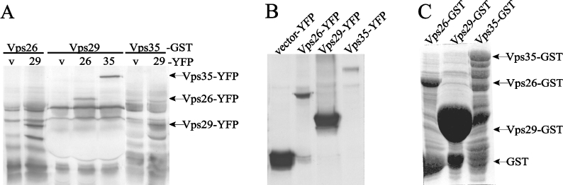
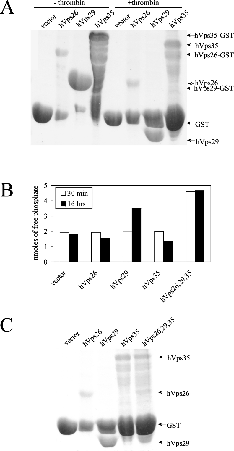
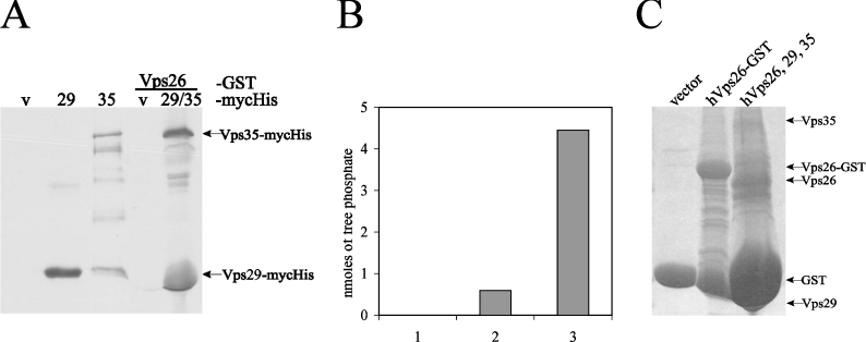
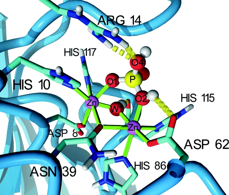
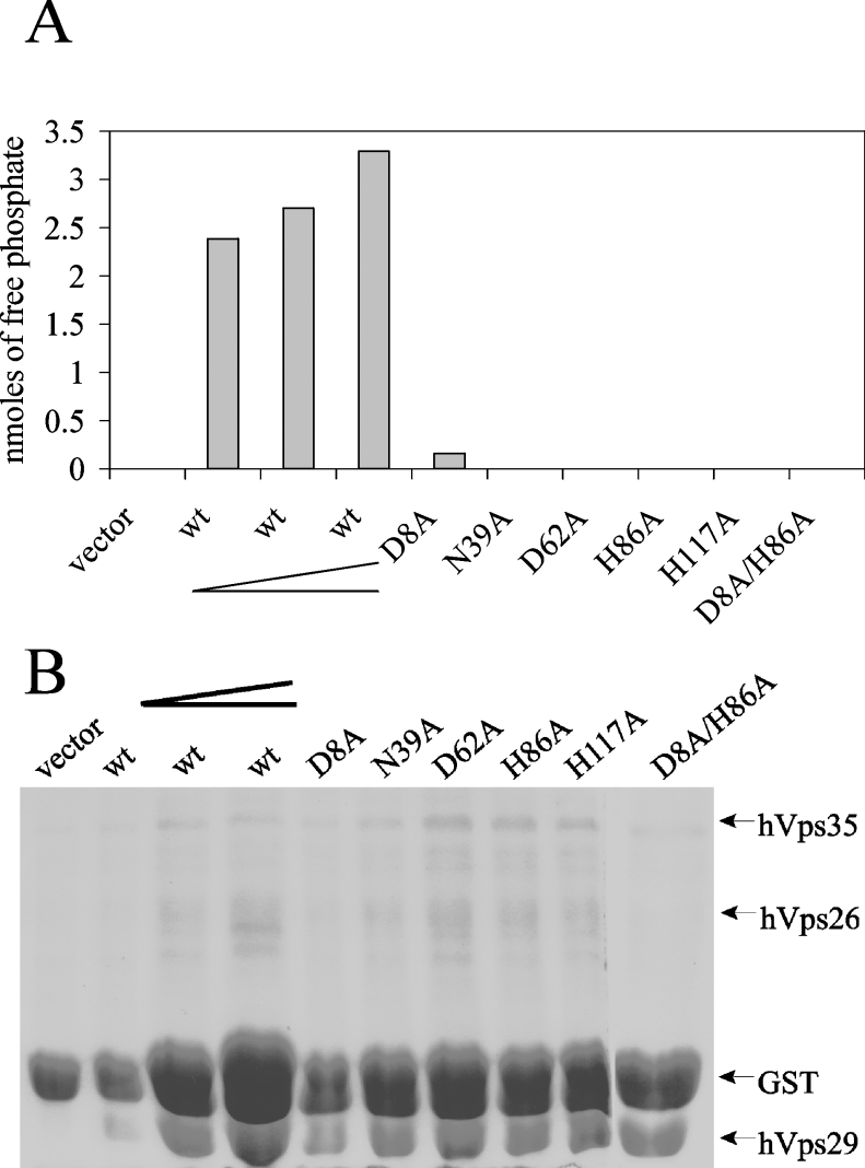
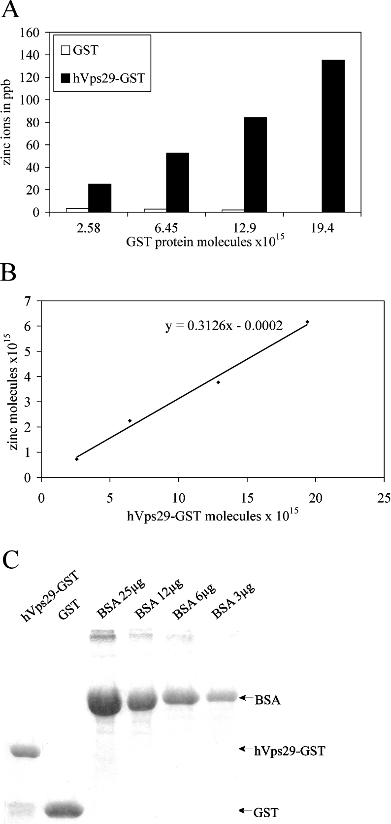
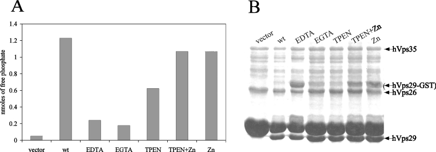
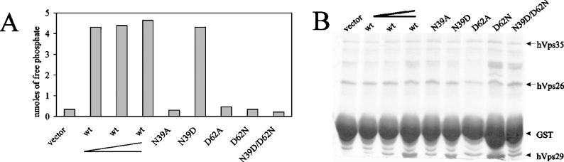
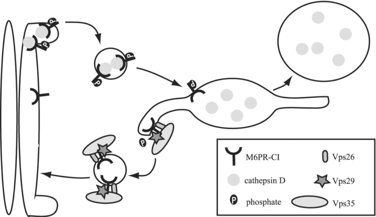
The retromer complex is involved in the retrograde transport of the CI-M6PR (cation-independent mannose 6-phosphate receptor) from endosomes to the Golgi. It is a hetero-trimeric complex composed of Vps26 (vacuolar sorting protein 26), Vps29 and Vps35 proteins, which are conserved in eukaryote evolution. Recently, elucidation of the crystal structure of Vps29 revealed that Vps29 contains a metallo-phosphoesterase fold [Wang, Guo, Liang, Fan, Zhu, Zang, Zhu, Li, Teng, Niu et al. (2005) J. Biol. Chem. 280, 22962-22967; Collins, Skinner, Watson, Seaman and Owen (2005) Nat. Struct. Mol. Biol. 12, 594-602]. We demonstrate that recombinant hVps29 (human Vps29) displays in vitro phosphatase activity towards a serine-phosphorylated peptide, containing the acidic-cluster dileucine motif of the cytoplasmatic tail of the CI-M6PR. Efficient dephosphorylation required the additional presence of recombinant hVps26 and hVps35 proteins, which interact with hVps29. Phosphatase activity of hVps29 was greatly decreased by alanine substitutions of active-site residues that are predicted to co-ordinate metal ions. Using inductively coupled plasma MS, we demonstrate that recombinant hVps29 binds zinc. Moreover, hVps29-dependent phosphatase activity is greatly reduced by non-specific and zinc-specific metal ion chelators, which can be completely restored by addition of excess ZnCl2. The binuclear Zn2+ centre and phosphate group were modelled into the hVps29 catalytic site and pKa calculations provided further insight into the molecular mechanisms of Vps29 phosphatase activity. We conclude that the retromer complex displays Vps29-dependent in vitro phosphatase activity towards a serinephosphorylated acidic-cluster dileucine motif that is involved in endosomal trafficking of the CI-M6PR. The potential significance of these findings with respect to regulation of transport of cycling trans-Golgi network proteins is discussed.


Figures

Figure 1
Figure 1. Interaction of hVps29 with hVps26 and hVps35 in vitro
(A) GST fusion proteins were used as an affinity matrix for hVps26, hVps29 and hVps35 YFP fusion proteins present in the lysates of transiently transfected NIH3T3 cells. Affinity precipitated proteins were visualized by Western-blot analysis with anti-GFP antibodies (v: vector; 26: Vps26; 29: Vps29; and 35: Vps35). (B) Western blot of whole cell lysate samples of NIH3T3 cells transfected with the YFP fusion constructs, detected with anti-GFP antibodies. (C) Coomassie Blue staining of the GST fusion constructs used for the pull-down assay.
Figure 2
Figure 2. Phosphatase activity of hVps29 on a CI-M6PR phosphopeptide
(A) Coomassie Blue staining of recombinantly expressed GST fusion proteins of vector (26 kDa), hVps26 (36 kDa), hVps29 (20 kDa) and hVps35 (87 kDa) before and after thrombin cleavage. (B) Detected amounts of free phosphate in an in vitro phosphatase assay on the SFHDDpSDEDLLHI phosphopeptide using recombinantly expressed vector, hVps26, hVps29 or hVps35 or a mixture of these three proteins. Levels of free phosphate were determined after 30 min and after 16 h of incubation at 37 °C. (C) Coomassie Blue staining of the remaining supernatant of the protein samples used for the in vitro phosphatase assay.
Figure 3
Figure 3. Phosphatase assay on affinity-precipitated retromer complex
(A) Western blot with anti-c-Myc antibody. Lanes 1–3, expression of purified Myc–His constructs following MCAC with Ni-NTA beads. Lanes 4–5, hVps26–GST affinity precipitation following incubation with Myc–His vector or with purified Myc–His hVps29 and hVps35 protein (v: vector; 29: Vps29; and 35: Vps35). (B) Detected amounts of free phosphate in an in vitro phosphatase assay on the SFHDDpSDEDLLHI phosphopeptide using GST vector (bar 1), an affinity precipitation of Vps26–GST containing Vps29 and Vps35 Myc–His fusion proteins (bar 2) or as a positive control, a mixture of hVps26, hVps29 and hVps35 proteins (bar 3), as used in Figure 2. Levels of free phosphate were determined after 30 min of incubation. (C) Coomassie Blue staining of the remaining supernatant of the protein samples used for the in vitro phosphatase assay.
Figure 4
Figure 4. Molecular model of the hVps29 active site
The catalytic site of hVps29 showing two Zn2+ ions, the co-ordinating amino acid side chains and a bound phosphate. The hydroxide ion bridging the two metal ions is labelled ‘W1’ and is hydrogen-bonded to the backbone oxygen of His115. Only polar hydrogen atoms are shown. The serine residue of the substrate peptide has been omitted, but should be connected to the now protonated phosphate oxygen. The image was created with YASARA (http://www.yasara.org).
Figure 5
Figure 5. Inhibition of the phosphatase activity of hVps29 by catalytic-site mutants
(A) Detected amounts of free phosphate in an in vitro phosphatase assay on the SFHDDpSDEDLLHI phosphopeptide using recombinantly expressed hVps26 and hVps35 in combination with vector, wild-type hVps29 in increasing concentrations, as indicated by the elongated chevron, or hVps29 mutants D8A, N39A, D62A, H86A, H117A or D8A/H86A. Levels of free phosphate were determined after 30 min of incubation. (B) Coomassie Blue staining of the remaining supernatant of the protein samples used for the in vitro phosphatase assay.
Figure 6
Figure 6. Detected amounts of zinc ions in hVps29–GST by ICP-MS
(A) Binding of zinc ions (in ppb) to increasing amounts of recombinantly expressed hVps29–GST fusion protein or GST alone. (B) Linear increase in the amount of zinc induced by increasing amounts of recombinantly expressed hVps29–GST fusion protein corrected for binding to GST alone. The slope of the line indicates the molar ratio of hVps29–GST/zinc. (C) Coomassie Blue staining of a small amount of the recombinantly expressed proteins used in the ICP-MS analysis. The protein amount was measured by photospectrometric analysis (results not shown), and also by comparison with BSA levels.
Figure 7
Figure 7. Inhibition of the phosphatase activity of hVps29 by metal ion chelators
(A) Detected amounts of free phosphate in an in vitro phosphatase assay on the SFHDDpSDEDLLHI phosphopeptide using recombinantly expressed hVps26 and hVps35 in combination with vector as a control or wild-type hVps29. Samples were treated with 5 mM of the indicated metal ion chelators prior to thrombin cleavage. When indicated, ZnCl2 was added to the reaction mixture to a final concentration of 1 mM. Levels of free phosphate were determined after 30 min of incubation at 37 °C. (B) Coomassie Blue staining of the remaining supernatant of the protein samples used for the in vitro phosphatase assay.
Figure 8
Figure 8. Effect of the Asn39/Asp62 pair on the phosphatase activity
(A) Detected amounts of free phosphate in an in vitro phosphatase assay on the SFHDDpSDEDLLHI phosphopeptide using recombinantly expressed hVps26 and hVps35 in combination with vector, wild-type hVps29 in increasing concentrations, as indicated by the elongated chevron, and hVps29 mutants N39A, N39D, D62A, D62N and N39D/D62N. Levels of free phosphate were determined after 30 min of incubation at 37 °C. (B) Coomassie Blue staining of the remaining supernatant of the protein samples used for the in vitro phosphatase assay.
Figure 9
Figure 9. Model of phosphate-dependent transport of the CI-M6PR
The CI-M6PR binds cathepsin D in the TGN. Binding of GGAs to the phosphorylated CI-M6PR causes the transport of the ligand–receptor complex to the endosome. In the endosome, the ligand dissociates from the receptor and after binding to Vps35, Vps29 is able to dephosphorylate the receptor. The retromer recycles the CI-M6PR to the TGN.

References

    1. Bankaitis V. A., Johnson L. M., Emr S. D. Isolation of yeast mutants defective in protein targeting to the vacuole. Proc. Natl. Acad. Sci. U.S.A. 1986;83:9075–9079. - PMC - PubMed
    1. Robinson J. S., Klionsky D. J., Banta L. M., Emr S. D. Protein sorting in Saccharomyces cerevisiae: isolation of mutants defective in the delivery and processing of multiple vacuolar hydrolases. Mol. Cell. Biol. 1988;8:4936–4948. - PMC - PubMed
    1. Rothman J. H., Stevens T. H. Protein sorting in yeast: mutants defective in vacuole biogenesis mislocalize vacuolar proteins into the late secretory pathway. Cell. 1986;47:1041–1051. - PubMed
    1. Arighi C. N., Hartnell L. M., Aguilar R. C., Haft C. R., Bonifacino J. S. Role of the mammalian retromer in sorting of the cation-independent mannose 6-phosphate receptor. J. Cell Biol. 2004;165:123–133. - PMC - PubMed
    1. Collins B. M., Skinner C. F., Watson P. J., Seaman M. N., Owen D. J. Vps29 has a phosphoesterase fold that acts as a protein interaction scaffold for retromer assembly. Nat. Struct. Mol. Biol. 2005;12:594–602. - PubMed

Publication types