

Review
doi: 10.1186/gb-2006-7-5-216.
Epub 2006 May 30.
An overview of the serpin superfamily
Affiliations
- PMID: 16737556
- PMCID: PMC1779521
- DOI: 10.1186/gb-2006-7-5-216
Item in Clipboard
Review
An overview of the serpin superfamily
Genome Biol.
2006.
Abstract
Serpins are a broadly distributed family of protease inhibitors that use a conformational change to inhibit target enzymes. They are central in controlling many important proteolytic cascades, including the mammalian coagulation pathways. Serpins are conformationally labile and many of the disease-linked mutations of serpins result in misfolding or in pathogenic, inactive polymers.
Figures
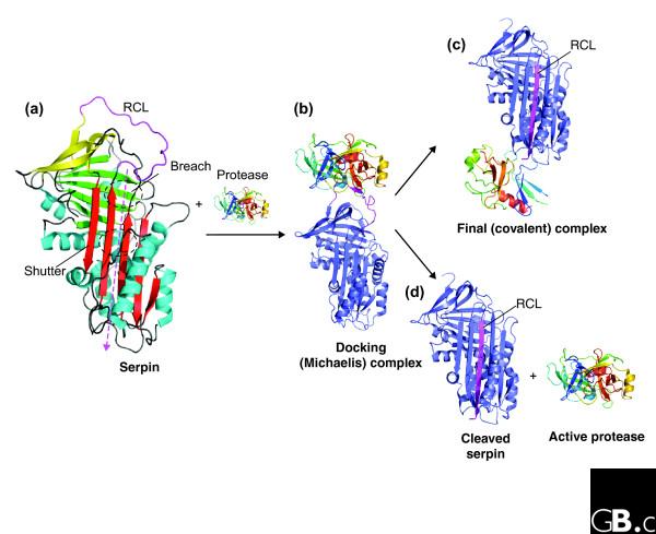
The structure and mechanism of inhibitory serpins. (a) The structure of native SERPINA1 (Protein Data Bank (PDB) code 1QLP) [32]. The A sheet is in red, the B sheet in green and the C sheet in yellow; helices (hA-hI) are in blue. The reactive center loop (RCL) is at the top of the molecule, in magenta. The position of the breach and the shutter are labeled and the path of RCL insertion indicated (magenta dashed line). Both of these regions contain several highly conserved residues, many of which are mutated in various serpinopathies. (b) The Michaelis or docking complex between SERPINA1 and inactive trypsin (PDB code 1OPH) [36], with the protease (multicolors) docked onto the RCL (magenta). Upon docking with an active protease (b), two possible pathways are apparent. (c) The final serpin enzyme complex (PDB code 1EZX [12]). The serpin has undergone the S to R transition, and the protease hangs distorted at the base of the molecule. (d) The structure of cleaved SERPINA1 is shown (PDB code 7API) [93]) with the RCL (magenta) forming the fourth strand of β-sheet A. The result of serpin substrate-like behavior can be seen where the protease has escaped the conformational trap, leaving active protease and inactive, cleaved serpin. Certain serpin mutations, particularly non-conservative substitutions within the hinge region of the RCL, result in substrate-like, rather than inhibitory, behavior [94].
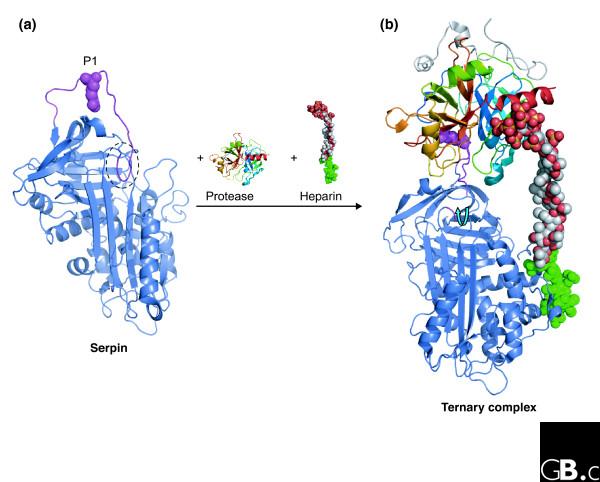
Modulation of serpin conformation by cofactors. (a) The structure of native SERPINC1 (PDB code 2ANT) [95]. The partial insertion of the RCL (two residues) into the top of β-sheet A is circled, and the position of the P1 residue is shown (magenta spheres). (b) The structure of the ternary complex between SERPINC1, inactive thrombin (the Ser195Ala mutant) and a synthetic long-chain heparin construct (PDB code 1TB6) [43]. A specific high-affinity pentasaccharide (green) on the heparin interacts with the heparin-binding site on SERPINC1 (on and around helix hD) and promotes expulsion of the RCL (blue arrow) and rearrangement of the P1 residue (magenta spheres).
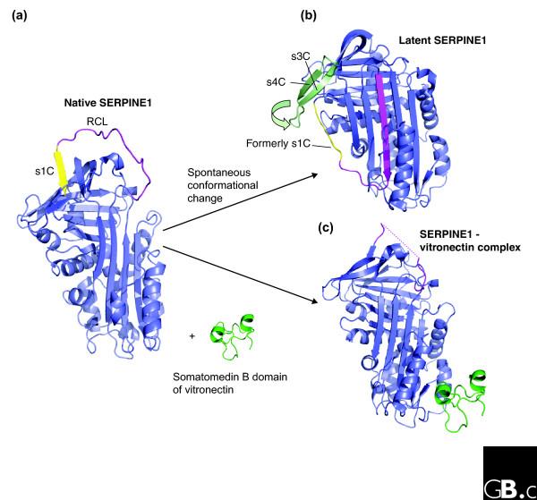
Spontaneous conformational change in serpins. (a) Structure of native SERPINE1 (PDB code 1B3K) [96]. The RCL is in magenta and strand s1c of β-sheet C is in yellow. (b) The structure of latent SERPINE1 (PDB code 1DVN) [53,97], which can form by spontaneous conversion from the native protein. The RCL (magenta) is inserted into β-sheet A. In order to enable full insertion of the RCL, s1C of β-sheet C (pale yellow) has peeled off. In addition, conformational change in the strands s3C and s4C (pale green) is indicated. (c) Structure of SERPINE1 (blue) in complex with the somatomedin B domain (green) of vitronectin (PDB code 1OC0) [54]. The interaction with vitronectin locks SERPINE1 in the native, active conformation.
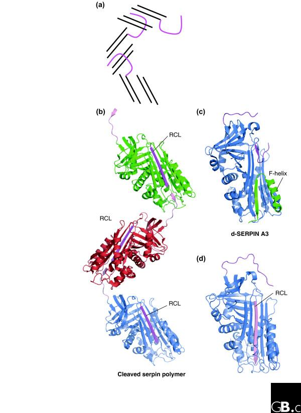
Structure of serpin polymers and other inactive conformers. (a) Schematic diagram of domain swapping in serpins; the RCL of one molecule (magenta loop), is docked into β-sheet A (black lines) of the next (only four strands of β-sheet A are shown). (b) Structure of a cleaved serpin polymer (PDB code 1D5S) [57], showing the promiscuous nature of the RCL. Cleavage at the P5/P6 position has resulted in RCL (magenta) insertion into β-sheet A; the 'gap' at the bottom of β-sheet A is filled with the P5-P1 portion (pale pink) from an RCL from another molecule. (c) The structure of an alternative confirmation of SERPINA3 -δ-SERPINA3 (PDB code 1QMN) [62]. Four residues of the RCL (magenta) are inserted into the top of β-sheet A. The F-helix (green) has partially unwound and filled the bottom half of β-sheet A. (d) Serpins can accept a peptide with the sequence of the RCL (pale pink) into β-sheet A (PDB code 1BR8) [98].
References
-
- Silverman GA, Bird PI, Carrell RW, Church FC, Coughlin PB, Gettins PG, Irving JA, Lomas DA, Luke CJ, Moyer RW, et al. The serpins are an expanding superfamily of structurally similar but functionally diverse proteins. Evolution, mechanism of inhibition, novel functions, and a revised nomenclature. J Biol Chem. 2001;276:33293–33296. doi: 10.1074/jbc.R100016200. - DOI - PubMed
-
- Irving JA, Steenbakkers PJ, Lesk AM, Op den Camp HJ, Pike RN, Whisstock JC. Serpins in prokaryotes. Mol Biol Evol. 2002;19:1881–1890. - PubMed
Publication types
MeSH terms
Substances
LinkOut - more resources
Full Text Sources
Other Literature Sources
