. 2006 Aug 4;281(31):22289-22298.
doi: 10.1074/jbc.M602664200. Epub 2006 May 31.

Autosomal recessive retinitis pigmentosa and E150K mutation in the opsin gene

Affiliations

Autosomal recessive retinitis pigmentosa and E150K mutation in the opsin gene

Li Zhu et al. J Biol Chem. .

Abstract

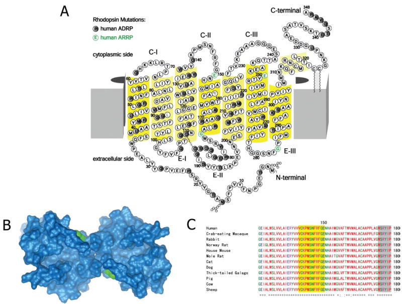
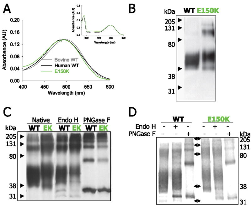
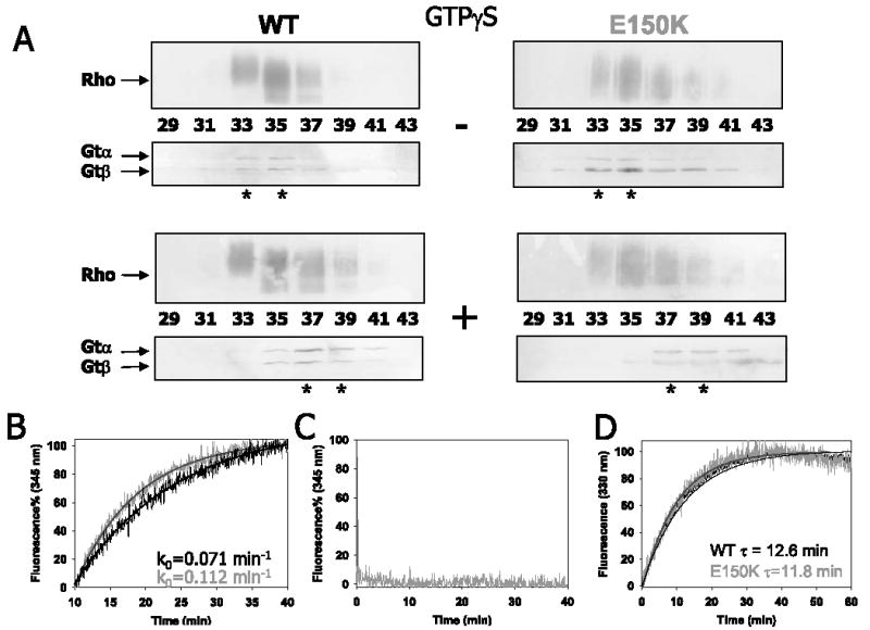
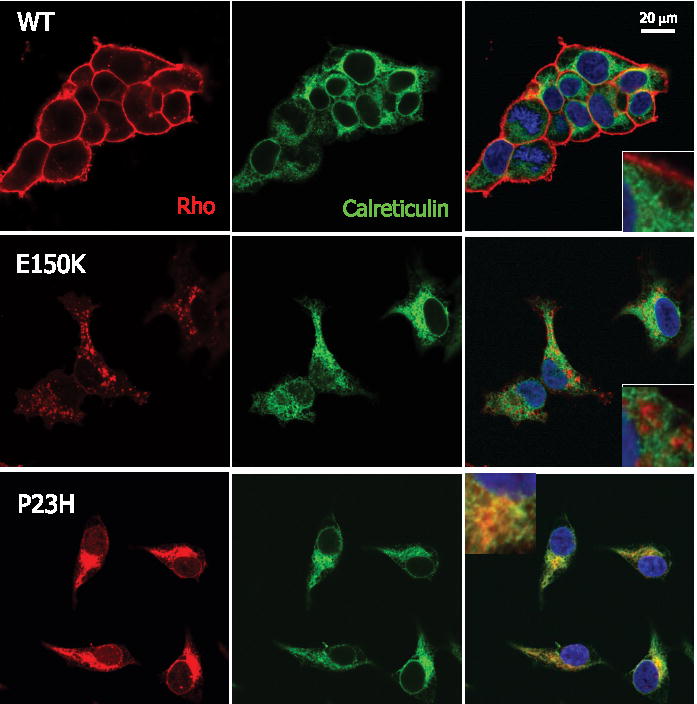
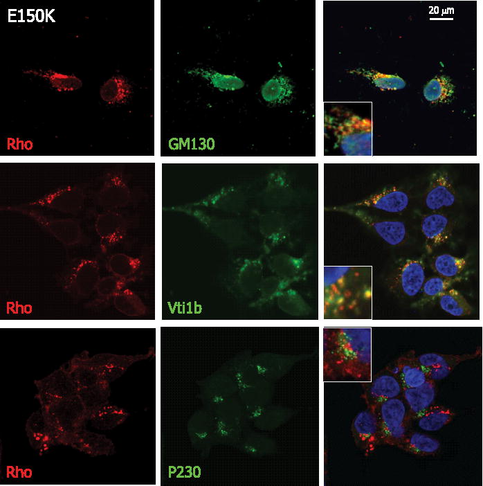
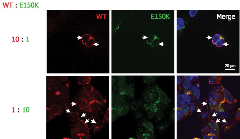
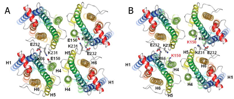
Retinitis pigmentosa (RP) is a heterogeneous group of hereditary disorders of the retina caused by mutation in genes of the photoreceptor proteins with an autosomal dominant (adRP), autosomal recessive (arRP), or X-linked pattern of inheritance. Although there are over 100 identified mutations in the opsin gene associated with RP, only a few of them are inherited with the arRP pattern. E150K is the first reported missense mutation associated with arRP. This opsin mutation is located in the second cytoplasmic loop of this G protein-coupled receptor. E150K opsin expressed in HEK293 cells and reconstituted with 11-cis-retinal displayed an absorption spectrum similar to the wild type (WT) counterpart and activated G protein transducin slightly faster than WT receptor. However, the majority of E150K opsin showed a higher apparent molecular mass in SDS-PAGE and was resistant to endoglycosidase H deglycosidase. Instead of being transported to the plasma membrane, E150K opsin is partially colocalized with the cis/medial Golgi compartment markers such as GM130 and Vti1b but not with the trans-Golgi network. In contrast to the endoplasmic reticulum-retained adRP mutant, P23H opsin, Golgi-retained E150K opsin did not influence the proper transport of the WT opsin when coexpressed in HEK293 cells. This result is consistent with the recessive pattern of inheritance of this mutation. Thus, our study reveals a novel molecular mechanism for retinal degeneration that results from deficient export of opsin from the Golgi apparatus.


Figures

FIGURE 1
FIGURE 1. E150K and other RP mutations in opsin
A, the two-dimensional model of opsin with indicated nonsense/missense mutations related to arRP (in green) and adRP (in black). B, three-dimensional model of E150K Rho with the cytoplasmic side facing outside and the mutation in green. C, the amino acid sequence alignment of 12 mammalian opsins around Glu150 (residues 121–180). The yellow background indicates the cytoplasmic loop, the white background indicates the transmembrane helices, and the gray background indicates the intradiscal loop.
FIGURE 2
FIGURE 2. Spectra, SDS-PAGE migration, and deglycosylation of E150K Rho
A, the visible absorbance spectra of immunoaffinity-purified bovine (gray, 498 nm), human (black, 496 nm) WT, and human E150K mutant (green, 496 nm) Rho. Inset, an extended UV-visible range of the spectrum. B, silver staining of the SDS-PAGE gel of immunoaffinity-purified WT opsin on the left and E150K opsin on the right. The molecular mass is shown by the arrows on the left side (kDa). The results of deglycosylation are shown in C and D. WT and E150K opsins were deglycosylated by either Endo H or PNGase F as indicated. C shows silver staining of the native or deglycosylated WT/E150K opsin as marked. D shows the immunoblot of deglycosylated WT/E150K opsin probed with anti-Rho 1D4 antibody. Molecular mass markers (in kDa) are 205, 131, 80, 38, and 31.
FIGURE 3
FIGURE 3. Gt binding, activation, and Meta II decay
A, Gt binding assay of WT/E150K Rho by size exclusion chromatography. Immunoblots are shown using anti-Rho (top panels), and the anti-Gtα or anti-Gtβ (bottom panels), with or without the addition of GTPγS. The number of fractions is listed. B, Gt activation by WT or E150K Rho monitored by the increase in fluorescence at 345 nm. The initial rates calculated as a pseudo first order reaction for WT and E150K are 0.071 and 0.112 min−1, respectively. C, E150K opsin showed no constitutive Gt activation. D, Meta II decay of WT/E150K measured by fluorescence emission at 330 nm. The absolute fluorescence intensity changes were comparable (in arbitrary units) 82– 86 for WT Rho and 85– 89 for the E150K mutant. The relaxation time (τ) for WT and E150K Rho is 13.6 and 11.8 min, respectively.
FIGURE 4
FIGURE 4. Chemical cross-linking of E150K opsin
Immunoblots of purified cross-linked WT or E150K opsins deglycosylated with PNGase F. WT or E150K opsins were cross-linked in the membranes before immunoaffinity purification. The corresponding samples in lanes 1 and 2 are reduced in lanes 3 and 4 by 100 mM DTT for 5 min before SDS-PAGE.
FIGURE 5
FIGURE 5. Intracellular colocalization of opsin with calreticulin, an ER marker
HEK293 cells expressing WT (top row), E150K (middle row), or P23H (bottom row) opsin are stained with both anti-Rho 1D4 (red) and anti-calreticulin (green). The nuclei are stained by Hoechst 33342 (blue). The right column shows the merged images. The insets in the right column are high magnification images of the merged images. The scale bar represents 20 μ m.
FIGURE 6
FIGURE 6. Intracellular colocalization of E150K opsin with Golgi markers
From top to bottom, anti-Rho 1D4 is used together with anti-GM130 (cis), anti-Vti1b (cis/medial), or anti-P230 (trans) to examine the intracellular colocalization of E150K opsin (red) and the Golgi apparatus (green).
FIGURE 7
FIGURE 7. Cotransfection of WT and E150K opsins
Different ratios of WT and E150K construct, 10/1 and 1/10, respectively, were used to cotransfect HEK293 cells. WT opsin was labeled with Cy3-conjugated anti-FLAG (red) antibody, and E150K opsin was labeled with fluorescein isothiocyanate-conjugated anti-c-Myc antibody (green). The nuclei were stained by Hoechst 33342 dye (blue). The bar represents 20 μm. The arrows indicate the plasma membrane expression of the opsins.
FIGURE 8
FIGURE 8. Molecular modeling of Rho tetramer in the membrane
View from cytoplasmic side. Rho dimers are located horizontally, so mutation does not prevent formation of dimers (interface H4-H5) but rather longer oligomers. A, molecular model of WT Rho tetramer. B, molecular model of E150K Rho tetramer.

References

    1. Dryja TP, Li T. Hum Mol Genet. 1995;4:1739–1743. - PubMed
    1. Filipek S, Stenkamp RE, Teller DC, Palczewski K. Annu Rev Physiol. 2003;65:851–879. - PMC - PubMed
    1. Palczewski K. Annu Rev Biochem. 2005;75:743–767. - PMC - PubMed
    1. Rosenfeld PJ, Cowley GS, McGee TL, Sandberg MA, Berson EL, Dryja TP. Nat Genet. 1992;1:209–213. - PubMed
    1. Ridge KD, Lee SS, Abdulaev NG. J Biol Chem. 1996;271:7860–7867. - PubMed

Publication types