. 2006 Jun 6;103(23):8703-8.
doi: 10.1073/pnas.0602569103. Epub 2006 May 31.

Mutant nuclear lamin A leads to progressive alterations of epigenetic control in premature aging

Affiliations

Mutant nuclear lamin A leads to progressive alterations of epigenetic control in premature aging

Dale K Shumaker et al. Proc Natl Acad Sci U S A. .

Abstract

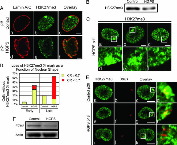
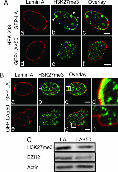
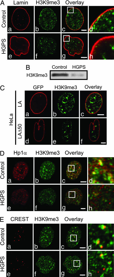
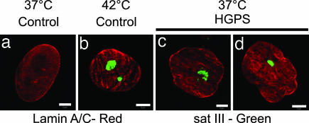
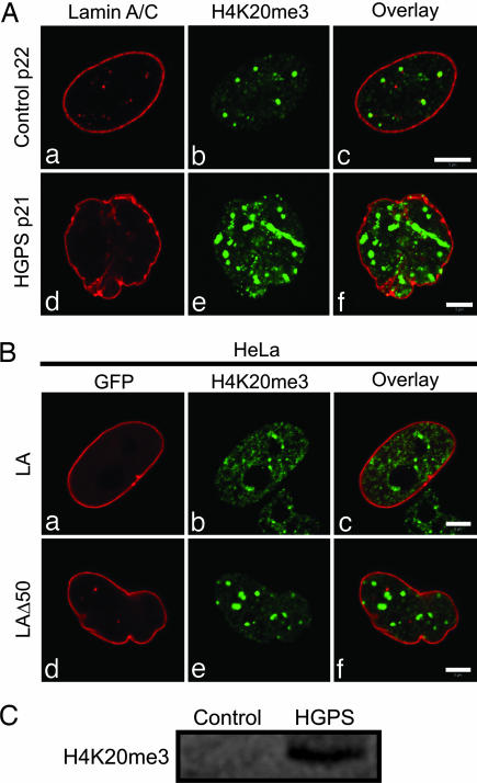
The premature aging disease Hutchinson-Gilford Progeria Syndrome (HGPS) is caused by a mutant lamin A (LADelta50). Nuclei in cells expressing LADelta50 are abnormally shaped and display a loss of heterochromatin. To determine the mechanisms responsible for the loss of heterochromatin, epigenetic marks regulating either facultative or constitutive heterochromatin were examined. In cells from a female HGPS patient, histone H3 trimethylated on lysine 27 (H3K27me3), a mark for facultative heterochromatin, is lost on the inactive X chromosome (Xi). The methyltransferase responsible for this mark, EZH2, is also down-regulated. These alterations are detectable before the changes in nuclear shape that are considered to be the pathological hallmarks of HGPS cells. The results also show a down-regulation of the pericentric constitutive heterochromatin mark, histone H3 trimethylated on lysine 9, and an altered association of this mark with heterochromatin protein 1alpha (Hp1alpha) and the CREST antigen. This loss of constitutive heterochromatin is accompanied by an up-regulation of pericentric satellite III repeat transcripts. In contrast to these decreases in histone H3 methylation states, there is an increase in the trimethylation of histone H4K20, an epigenetic mark for constitutive heterochromatin. Expression of LADelta50 in normal cells induces changes in histone methylation patterns similar to those seen in HGPS cells. The epigenetic changes described most likely represent molecular mechanisms responsible for the rapid progression of premature aging in HGPS patients.


Conflict of interest statement

Conflict of interest statement: No conflicts declared.

Figures

Fig. 1.
Fig. 1.
H3K27me3 decreases and XIST RNA remains with Xi at all passages in HGPS cells. (A) Cells were processed for immunofluorescence with antibodies against LA/C (a and d) and H3K27me3 (b and e; overlay, c and f). Control cell nuclei at p9 displayed a distinct LA/C nuclear rim (a) associated with a compact Xi (b). The HGPS cells showed a change in nuclear morphology by p21 by using anti-LA/C (d), and there was no obvious Xi revealed by anti-H3K27me3 (e). (Scale bars, 5 μm.) (B) There was a decrease in H3K27me3 by Western blot analysis of HGPS compared with control total cell extracts at p20. (C) In early-passage HGPS cells stained with anti-H3K27me3, the Xi appeared either as a uniformly stained compact domain (a and d) or a loose array of closely spaced granules (b and e), or it could not be detected (a typical lamina region in a nucleus with no Xi staining is depicted in the white box; c and f). d–f are enlargements (×4) of the regions in the white boxes in a–c. (Scale bars, 5 μm.) (D) Nuclear contour ratios (CR; see also ref. 1) were determined for control and HGPS cells at early and late passages (nuclei with a CR ≥ 0.7 were considered nonlobulated, and those with a CR < 0.7 were considered lobulated). In early-passage HGPS cells, ≈43% of the nuclei lost their H3K27me3 Xi mark, and ≈80% of these were normally shaped. In late-passage cells, ≈63% lost this mark, whereas ≈20% of these nuclei were normally shaped. In early-passage controls, ≈6% of the nuclei did not contain an obvious Xi, as distinguished by anti-H3K27me3. At late passages, this number increased to ≈20%. (E) Control (a–d) and HGPS cells (e–l) were prepared for XIST RNA FISH and stained with anti-H3K27me3. (a–d) In controls at early and late passages (p13–20), XIST RNA was associated with the Xi, and ≈93% (n = 102) also contained the H3K27me3 mark. (e–l) In HGPS cells, XIST FISH revealed an Xi at all passages, whereas by p17–18, the H3K27me3 staining on Xi was lost (≈53%; n = 82). (i–l) In lobulated HGPS nuclei lacking the H3K27me3 mark on Xi, the XIST RNA staining was more dispersed. d, h, and l are enlargements (×5.6) of the overlay regions in the white boxes (c, g, and k). (Scale bars, 10 μm.) (F) Western blotting shows a decrease in EZH2 in HGPS cells compared with controls at p14. Actin was used as a loading control.
Fig. 2.
Fig. 2.
Expression of GFP-LAΔ50 in HEK293 and HeLa cells. (A) HEK293 cells transiently expressing either GFP-LA or GFP-LAΔ50 were stained with anti-H3K27me3. In controls (GFP-LA), the lamina appeared normal (a), and H3K27me3 staining revealed several Xi (arrowheads, b) and arrays of peripheral heterochromatin (∗, b) colocalizing with the lamina (c). Most cells expressing GFP-LAΔ50 showed a dramatic loss of Xi and peripheral heterochromatin staining with anti-H3K27me3, even in those retaining normal nuclear shapes (df). (B) HeLa cells expressing either GFP-LA (a–d) or GFP-LAΔ50 (e–h) were stained with anti-H3K27me3. GFP-LA-expressing cells displayed a normally shaped nucleus (a) and punctate H3K27me3 staining throughout the nucleus, with prominent arrays at the nuclear periphery (∗, b), where it overlaps with the lamina (c and d). Expression of GFP-LAΔ50 caused lobulations in the nuclear envelopes of the majority of HeLa cells (e), and there was an overall loss of H3K27me3 staining, especially in the region of the lamina (f–h). Enlargements (×7.4) of the overlay areas indicated by white boxes (c and g) are shown (d and h). (C) Whole-cell extracts derived from HeLa cells expressing GFP-LA or GFP-LAΔ50 for 24–48 h were analyzed by immunoblotting. Note the reductions in H3K27me3 and EZH2 in cells expressing the mutant protein. Actin was used as a loading control. (Scale bars, 5 μm.)
Fig. 3.
Fig. 3.
LAΔ50 expression results in changes in constitutive heterochromatin. (A) HGPS and control cells were stained by using antibodies directed against LA/C and H3K9me3. (a–d) Early- and late-passage control cell nuclei appeared normal. The H3K9me3 staining pattern consisted of small foci dispersed throughout the nucleoplasm and in the lamina region. (e–h) In late-passage (p21) HGPS cells, the H3K9me3 immunofluorescence pattern was altered in lobulated nuclei. These nuclei showed an overall decrease in the amount of H3K9me3 staining, which was most obvious in the lamina region. Enlargements (×4.5) of the overlay areas indicated by white boxes (c and g) are shown (d and h). (B) There is a decrease in the amount of H3K9me3 in HGPS cells (p23) compared with control cells (p24), as indicated by Western blotting. (C) HeLa cells expressing either GFP-LA or GFP-LAΔ50 were stained with anti-H3K9me3. (a–c) In controls, the H3K9me3 staining pattern consisted of a few large foci interspersed among smaller foci throughout the nucleoplasm and in close proximity to the lamina. When GFP-LAΔ50 was expressed, the majority of HeLa cell nuclei were lobulated (d–f), and there were fewer H3K9me3 foci both in the nucleoplasm and the lamina (d–f). (D) Midpassage (p16) control and HGPS cells were stained with antibodies to H3K9me3 and Hp1α. Both antibodies stained large and small foci throughout the nucleoplasm. (a–d) In controls, many of the large foci stained with both antibodies, as seen in the overlay. (e–h) In HGPS cells, this colocalization is reduced. Enlargements (×4) of the overlay areas indicated by white boxes (c and g) are shown (d and h). (E) Control and HGPS cells were also double-labeled with CREST antiserum and anti-H3K9me3 (p16). (a–d) In controls, CREST revealed a typical pattern of kinetochores, and the majority of these were associated with H3K9me3. (e–h) In HGPS cells, many kinetochores were not associated with H3K9me3. Enlargements (×4.4) of the overlay areas indicated by white boxes (c and g) are shown (d and h). (Scale bars, 5 μm.)
Fig. 4.
Fig. 4.
Up-regulation of sat III transcripts in HGPS cells. Control and HGPS cells were analyzed by sat III RNA FISH. (a) No signal was observed in control cells grown at 37°C. (b) After heat shock, prominent stress bodies (green) were observed. (c and d) In HGPS cells grown at 37°C, stress bodies were observed in nuclei regardless of the extent of their lobulation. (Scale bars, 5 μm.)
Fig. 5.
Fig. 5.
Increased staining of H4K20me3 in late-passage HGPS cells. (A) Late-passage HGPS and control cells were stained with antibodies directed against LA/LC and H4K20me3. The fluorescence pattern looked similar in early and late passages (a; data not shown). (a–c) Nuclei of control cells at p22 displayed foci of H4K20me3 throughout the nucleoplasm that were frequently adjacent to lamin foci. In late-passage HGPS cells (p21), the H4K20me3 staining pattern consisted of much larger intensely staining structures. (d–f) There was also a loss of the association with lamin foci. (B a and d) HeLa cells expressing either GFP-LA or GFP-LAΔ50 were prepared for immunofluorescence by using anti-H4K20me3. (b and c) The H4K20me3 staining pattern consisted of mainly small foci throughout the nucleoplasm. (d–f) When GFP-LAΔ50 was expressed in HeLa cells, the majority of their nuclei were lobulated, and the H4K20me3 antibody showed large bright foci throughout the nucleoplasm. (C) Immunoblotting of control (p23) and HGPS cells (p24) for H4K20me3 showed a significant increase in HGPS cells. (Scale bars, 5 μm.)

References

    1. Goldman R. D., Shumaker D. K., Erdos M. R., Eriksson M., Goldman A. E., Gordon L. B., Gruenbaum Y., Khuon S., Mendez M., Varga R., Collins F. S. Proc. Natl. Acad. Sci. USA. 2004;101:8963–8968. - PMC - PubMed
    1. Hutchinson J. Medicochir. Trans. 1886;69:473–477. - PMC - PubMed
    1. Gilford M. Practitioner. 1904;73:188–217.
    1. Stables G. I., Morley W. N. J. R. Soc. Med. 1994;87:243–244. - PMC - PubMed
    1. de Sandre-Giovannoli A., Bernard R., Cau P., Navarro C., Amiel J., Boccaccio I., Lyonnet S., Stewart C. L., Munnich A., Le Merrer M., Levy N. Science. 2003;300:2055. - PubMed

Publication types

MeSH terms