. 2006 May 31;34(10):2887-905.
doi: 10.1093/nar/gkl295. Print 2006.

Identification of multiple distinct Snf2 subfamilies with conserved structural motifs

Affiliations

Identification of multiple distinct Snf2 subfamilies with conserved structural motifs

Andrew Flaus et al. Nucleic Acids Res. .

Abstract

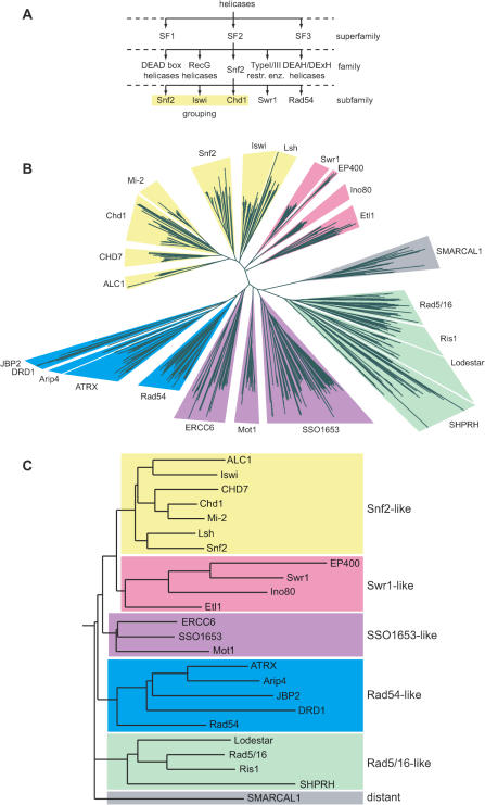
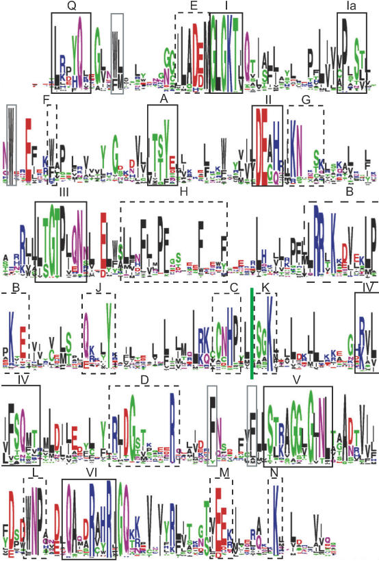
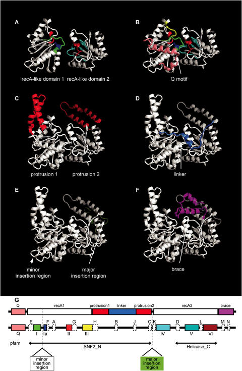
The Snf2 family of helicase-related proteins includes the catalytic subunits of ATP-dependent chromatin remodelling complexes found in all eukaryotes. These act to regulate the structure and dynamic properties of chromatin and so influence a broad range of nuclear processes. We have exploited progress in genome sequencing to assemble a comprehensive catalogue of over 1300 Snf2 family members. Multiple sequence alignment of the helicase-related regions enables 24 distinct subfamilies to be identified, a considerable expansion over earlier surveys. Where information is known, there is a good correlation between biological or biochemical function and these assignments, suggesting Snf2 family motor domains are tuned for specific tasks. Scanning of complete genomes reveals all eukaryotes contain members of multiple subfamilies, whereas they are less common and not ubiquitous in eubacteria or archaea. The large sample of Snf2 proteins enables additional distinguishing conserved sequence blocks within the helicase-like motor to be identified. The establishment of a phylogeny for Snf2 proteins provides an opportunity to make informed assignments of function, and the identification of conserved motifs provides a framework for understanding the mechanisms by which these proteins function.


Figures

Figure 1
Figure 1
Tree view of Snf2 family. (A) Schematic diagram illustrating hierarchical classification of superfamily, family and subfamily levels. (B) Unrooted radial neighbour-joining tree from a multiple alignment of helicase-like region sequences excluding insertions at the minor and major insertion regions from motifs I to Ia and conserved blocks C–K for 1306 Snf2 proteins identified in the Uniref database. The clear division into subfamilies is illustrated by wedge backgrounds, coloured by grouping of subfamilies. Subfamilies DRD1 and JBP2 were not clearly separated, as discussed in text. (C) In order to illustrate the relationship between subfamilies, a rooted tree was calculated using HMM profiles for full-length alignments of the helicase regions. Groupings of subfamilies are indicated by colouring as in (B).
Figure 2
Figure 2
Conserved residues within Snf2 helicase-like region. Sequence logo of global multiple alignment of 1306 Snf2 helicase-like region for alignment positions with residues in >90% of proteins. Helicase motifs are indicated in solid black boxes with roman numerals I–IV, additional conserved blocks are indicated in dashed black boxes with uppercase letters A–N, and conserved hydrophobic residues packing in the core of the structure by grey solid boxes. Motif and box labels as in Thoma et al. (47) with extensions. A comparison to other nomenclatures is in Supplementary Table S5. See Table 4 for actual distances between conserved blocks.
Figure 3
Figure 3
Conserved blocks contribute to distinctive structural features of Snf2 family proteins. Structural components of Snf2 family proteins relevant to the conservation are illustrated on the zebrafish Rad54A structure [pdb 1Z3I (153)]. (A) core recA-like domains 1 and 2 including colouring of helicase motifs (I in green, Ia in blue, II in bright red, III in yellow, IV in cyan, V in teal and VI in dark red). (B) Q motif (pink). (C) antiparallel alpha helical protrusions 1 and 2 (red) projecting from recA-like domains 1 and 2, respectively. (D) Linker spanning from protrusion 1 to protrusion 2 (middle blue). (E) Major insertion region behind protrusion 2 (light green). (F) triangular brace (magenta). (G) Schematic diagram showing location of structural elements and helicase motifs coloured as in A–F, with conserved blocks from Figure 2 shown as white boxes. Spans identified by Pfam profiles SNF2_N and Helicase_C are shown flanking the major insertion site.

References

    1. Gorbalenya A.E., Koonin E.V. Helicases: amino acid sequence comparisons and structure–function relationship. Curr. Opin. Struct. Biol. 1993;3:419–429.
    1. Gorbalenya A.E., Koonin E.V., Donchenko A.P., Blinov V.M. A novel superfamily of nucleoside triphosphate-binding motif containing proteins which are probably involved in duplex unwinding in DNA and RNA replication and recombination. FEBS Lett. 1988;235:16–24. - PMC - PubMed
    1. Pause A., Sonenberg N. Mutational analysis of a DEAD box RNA helicase: the mammalian translation initiation factor eIF-4A. EMBO J. 1992;11:2643–2654. - PMC - PubMed
    1. Tanner N.K., Cordin O., Banroques J., Doere M., Linder P. The Q motif: a newly identified motif in DEAD box helicases may regulate ATP binding and hydrolysis. Mol. Cell. 2003;11:127–138. - PubMed
    1. Subramanya H.S., Bird L.E., Brannigan J.A., Wigley D.B. Crystal structure of a DExx box DNA helicase. Nature. 1996;384:379–383. - PubMed

Publication types