. 2006 May 31;34(10):2966-75.
doi: 10.1093/nar/gkl357. Print 2006.

Polyadenylation of ribosomal RNA in human cells

Affiliations

Polyadenylation of ribosomal RNA in human cells

Shimyn Slomovic et al. Nucleic Acids Res. .

Abstract

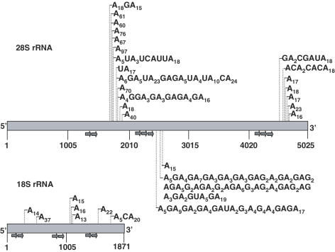
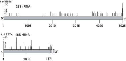
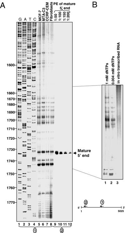
The addition of poly(A)-tails to RNA is a process common to almost all organisms. In eukaryotes, stable poly(A)-tails, important for mRNA stability and translation initiation, are added to the 3' ends of most nuclear-encoded mRNAs, but not to rRNAs. Contrarily, in prokaryotes and organelles, polyadenylation stimulates RNA degradation. Recently, polyadenylation of nuclear-encoded transcripts in yeast was reported to promote RNA degradation, demonstrating that polyadenylation can play a double-edged role for RNA of nuclear origin. Here we asked whether in human cells ribosomal RNA can undergo polyadenylation. Using both molecular and bioinformatic approaches, we detected non-abundant polyadenylated transcripts of the 18S and 28S rRNAs. Interestingly, many of the post-transcriptionally added tails were composed of heteropolymeric poly(A)-rich sequences containing the other nucleotides in addition to adenosine. These polyadenylated RNA fragments are most likely degradation intermediates, as primer extension (PE) analysis revealed the presence of distal fragmented molecules, some of which matched the polyadenylation sites of the proximal cleavage products revealed by oligo(dT) and circled RT-PCR. These results suggest the presence of a mechanism to degrade ribosomal RNAs in human cells, that possibly initiates with endonucleolytic cleavages and involves the addition of poly(A) or poly(A)-rich tails to truncated transcripts, similar to that which operates in prokaryotes and organelles.


Figures

Figure 1
Figure 1
Detection of 28S and 18S rRNA transcripts containing homopolymeric and heteropolymeric poly(A)-tails using oligo(dT) primed RT–PCR. The 28S and 18S rRNAs are schematically presented. The gene specific forward primers used for the PCR amplification and screening of the oligo(dT)-primed cDNAs are shown as arrows below the gene. Thin vertical lines indicate the positions of the added poly(A)-tail of each clone and the tail compositions are shown. The exact positions of the polyadenylated nucleotides, according to the gene sequence, are presented in the Supplementary Tables S3 and S4.
Figure 2
Figure 2
Detection of ESTs related to polyadenylated 28S and 18S rRNA transcripts. ESTs containing the 28S or 18S rRNAs with poly(A) or poly(A)-rich tails were identified by the bioinformatic tool, PolyAfinder. The points of addition of the poly(A) or poly(A)-rich tails are presented as thin vertical lines, while the length of the line indicates the number of ESTs which were polyadenylated at this location. Examples of the sequences of several heteropolymeric tails are presented in Table 1 and a full list, including the polyadenylation positions according to the gene sequence as well, is presented in Supplementary Tables S1 and S2 of the supplementary Data.
Figure 3
Figure 3
PE analysis of the 28S rRNA. (A) In an attempt to detect distal cleavage products, total RNA isolated from the cell types indicated at the top of lanes 5–8 was annealed to [32P] primer 28Sp1 and extended with reverse transcriptase. The resulting cDNA was fractionated on 7% denaturing PAGE along with a DNA sequencing ladder generated using a 28S rRNA DNA template and the same primer (lanes 1–4). The nucleotide numbers of the sequence of 28S rRNA are indicated to the left. In order to estimate the amount of the putative truncated transcripts, the mature 5′ end of the molecule was detected by PE with primer 28Sp5, located 99 nt downstream to the mature 28S rRNA 5′ end, using RNA purified from CCRF-CEM cells. Appropriated dilutions of this reaction, as shown at the top of lanes 9–12 were fractionated on the same gel. (B) Control reactions, in which RNA purified from CCRF-CEM cells was analyzed by PE in the presence of dNTPs at concentrations of 1 or 0.004 mM (lanes 1 and 2, respectively) in order to distinguish whether the observed signals were caused by modified nucleotides, are shown. Note that the significant signal located at nucleotide 1733 was not detected in this experiment. In lane 3, in vitro transcribed RNA corresponding to the analyzed region of 28S rRNA, was analyzed by PE to ensure that the signals did not arise from secondary structures that inhibit the reverse transcriptase.
Figure 4
Figure 4
cRT–PCR analysis detected additional 28S fragments which terminate in the region disclosed by oligo(dT) RT–PCR and PE. The combined results of the analysis of the 28S rRNA for truncated and polyadenylated fragments using cRT–PCR, oligo(dT) RT–PCR (dT-RT–PCR) and PE are schematically presented along the region of 1250–2100 nt. Horizontal lines represent the cDNAs obtained by each technique and are drawn to scale. The primers used for the PCR amplification and screening of the dT-RT–PCR and cRT–PCR cDNA, as well as the PE are shown by arrows. Details about the cRT–PCR clones are displayed in the Supplementary Table S5 and the clone # in the figure is related to that which appears in this table. The defined region of 1670 to 1780 that is enriched with 5′ and 3′ ends of detectable cleavage products is indicated with a gray background. Inset: A diagram explaining the experimental design. Following cleavage, the 28S rRNA is separated to proximal (upstream) and distal (downstream) fragments. PE discloses the 5′ end of the distal fragment while cRT–PCR and oligo(dT) RT–PCR analysis reveal the 3′ end of the proximal fragment, to which the poly(A) tail is added. Arrow heads indicate the primers used and dashed lines the cDNAs produced.

References

    1. Dreyfus M., Regnier P. The poly(A) tail of mRNAs: bodyguard in eukaryotes, scavenger in bacteria. Cell. 2002;111:611–613. - PubMed
    1. Kushner S.R. mRNA decay in prokaryotes and eukaryotes: different approaches to a similar problem. IUBMB Life. 2004;56:585–594. - PubMed
    1. Slomovic S., Portnoy V., Liveanu V., Schuster G. RNA polyadenylation in prokaryotes and organelles; different tails tell different tales. Crit. Rev. Plant Sci. 2006;25:65–77.
    1. Edmonds M. A history of poly(A) sequences: from formation to factors to function. Prog. Nucleic Acid Res. Mol. Biol. 2002;71:285–389. - PubMed
    1. Shatkin A.J., Manley J.L. The ends of the affair: capping and polyadenylation. Nature Struct. Biol. 2000;7:838–842. - PubMed

Publication types