. 2006 Jun;26(12):4499-510.
doi: 10.1128/MCB.00079-06.

Centromeric histone H3 is essential for vegetative cell division and for DNA elimination during conjugation in Tetrahymena thermophila

Affiliations

Centromeric histone H3 is essential for vegetative cell division and for DNA elimination during conjugation in Tetrahymena thermophila

Bowen Cui et al. Mol Cell Biol. 2006 Jun.

Abstract

The Tetrahymena thermophila CNA1 gene encodes the centromeric H3, Cna1p. Green fluorescent protein (GFP)-tagged Cna1p localizes in micronuclei in dots whose number and behavior during mitosis and conjugation are consistent with centromeres. During interphase, Cna1p-GFP localizes in peripheral dots, suggesting centromeres are associated with the nuclear envelope. Newly synthesized Cna1p-GFP enters micronuclei in mitosis and accumulates in the nucleoplasm. Its deposition at centromeres starts at early S phase and continues through most of S phase. CNA1 is required for vegetative cell growth. Knockdown of CNA1 genes in the somatic macronucleus results in micronuclear DNA loss and delayed chromosome segregation during mitosis. During conjugation, Cna1p-GFP disappears from the centromeres in the developing macronucleus, consistent with centromeric sequences being internal eliminated sequences. Surprisingly, zygotic CNA1 is required for efficient elimination of germ line-specific sequences during development of the new macronuclei but not for the RNA interference pathway, through which sequences are targeted for elimination. Zygotically expressed Cna1p localizes in the spherical structures in which the later stages of DNA elimination occur, and these structures cannot be formed in the absence of zygotic CNA1, suggesting that, in addition to functioning in centromeres, Cna1p may also play a role in organizing the formation of the DNA elimination structures.


Figures

FIG. 1.
FIG. 1.
Characterization of CNA1. (A) The schematic drawing shows that CenH3 has a histone fold domain (HFD) that shares high homology with H3 and an N-terminal region that is very diversified. (B) Centromere H3 proteins. tt, Tetrahymena thermophila; hs, Homo sapiens; mm, Mus musculus; dm, Drosophila melanogaster; sc, Saccharomyces cerevisiae. The sequences of Tetrahymena H3 (ttH3) and human H3 (hsH3) are included for comparison. The top panel shows the N-terminal regions of the proteins which have almost no homology. The bottom panel is the alignment of the HFDs of the proteins; the identity and similarity of a protein compared to ttCna1p are indicated. αN, α1, α2, and α3 indicate the α-helixes in the histone fold domains. The boxed area indicates the longer L1 loop in the CenH3s compared to those of H3s. (C) Expression pattern of the CNA1 gene. Total RNA from wild-type log-phase growing (G), starved (S), and conjugating cells (0, 2, 4, 6, 8, 10, 12, and 24 h postmixing) were used. The upper panel is the Northern blot probed with CNA1 coding sequence. The lower panel is the loading control of rRNA stained with ethidium bromide before blotting.
FIG. 2.
FIG. 2.
Localization of Cna1p-GFP protein in vegetative growing cells. (A) Diagram of the CNA1 GFP-tagged construct at the MTT1 locus. MTT1 coding sequence was replaced by CNA1-GFP coding sequence. The neo2 selectable marker was inserted into the 5′ flanking region of the MTT1 locus ∼900 bp upstream of the start codon. (B) Cna1p-GFP is localized as peripheral dots in MIC. Fifteen sequential cofocal images through a MIC with intervals of 0.28 μm, as well as the overlaid image (the last image in each panel), are shown. Scale bar, 2 μm. (C) Localization of Cna1p-GFP in log-phase cells. The first column is DAPI staining of the cells at lower magnification. Scale bar, 20 μm. The second (GFP) and third columns (merge of GFP and DAPI) are 4× magnifications of the boxed area indicated in the first column. Scale bars, 4 μm. Each picture shown is an overlaid image of eight sections through the nuclei obtained by confocal microscopy. a, interphase cell; b, MIC prometaphase; c, MIC metaphase; d, MIC anaphase; e, MIC telophase/start of S phase; f, MIC S phase, early MAC elongation; g and h, MIC S phase and MAC division; and i, interphase cell.
FIG. 3.
FIG. 3.
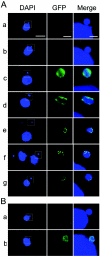
Cna1p-GFP deposition. The arrangement and magnifications are the same as those described in the legend to Fig. 2C. (A) Cna1p-GFP is deposited into centromeres during S phase. a, a growing MCG cell before induction of Cna1p-GFP expression; b to g, 2 h after induction; b, an unlabeled interphase cell; c and d, MIC M phase; e to g, MIC S phase. (B) Cna1p-GFP cannot be deposited into centromeres in starved cells in which the MICs are arrested at G2 phase. a, starved MCG cell before induction; b, 10 h after induction.
FIG. 4.
FIG. 4.
Localization of Cna1p in conjugating cells. (A) Localization of Cna1p-GFP. The first and second columns are GFP fluorescence and DAPI staining of the cells at lower magnification. Scale bars, 20 μm. The third (GFP) and fourth (merge of GFP and DAPI) columns are 4× magnifications of the boxed areas indicated in the second column. Scale bars, 4 μm. a, starved cell; b, early conjugation; c and d, crescent; e, early first meiotic division; f, first meiotic division; g, second meiotic division; h and i, prezygotic division; j, pronuclear exchange; k, nuclear fusion and first postzygotic division; l, anaphase of second postzygotic division; m, end of second postzygotic division; n, early MAC development; and o to t, late stages in MAC development. (B) Cna1p is localized in the DNA elimination structures. Wild-type cells were fixed at 13.5 h postmixing and were stained using anti-Cna1p antibody as described previously (6). Arrows indicate two DNA elimination structures in NM. Note that the Cna1p colocalizes with the DAPI-stained DNA elimination structures. Scale bar, 12 μm.
FIG. 5.
FIG. 5.
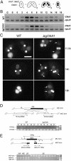
Macronuclear CNA1 gene is essential for vegetative growth and is required for proper chromosome segregation and MIC maintenance. (A) Diagram of the CNA1 knockout construct and the wild-type CNA1 locus. The CNA1 coding sequence is replaced by the neo2 cassette. The probe used for panel B is indicated. (B) Southern hybridization of the incomplete somatic CNA1 knockout strains (i-ΔsCNA1). Genomic DNA isolated from wild-type cells (WT) and i-ΔsCNA1 cells (assorted to the highest nonkilling concentration of paromomycin) was digested with ClaI and HincII and hybridized with the probe indicated in panel A. (C) Delayed chromosome segregation and small-MIC phenotype of i-ΔsCNA1 cells. Growing wild-type (WT) or i-ΔsCNA1 cells were fixed and stained with DAPI. Scale bar, 10 μm. Arrows indicate MICs.
FIG. 6.
FIG. 6.
DNA elimination is defective in conjugated germ line CNA1 knockout (ΔgCNA1) cells. (A) Schematic drawing of the mating process of ΔgCNA1 cells. In ΔgCNA1 heterokaryon cells, CNA1 genes are disrupted by the neo2 cassette in MIC but are wild type (WT) in MAC (1). When two such cells mate, they form a conjugating pair (2). The conjugating pair then develops to NM stage (3), when the old parental MAC stops transcription and starts degrading, and the new MACs, containing the same zygotic genome as the new MICs, start transcription. The conjugating pair then separates and gives rise to two exconjugants (4). (B) The zygotic CNA1 expression is up-regulated during late conjugation. The expression of the CNA1 gene was analyzed by reverse transcription-PCR, amplifying CNA1 and rpL21 (loading control) cDNAs at indicated time points. The up-regulation of CNA1 expression at 12 to 16 h in WT cells is not observed in ΔgCNA1 cells. (C) Endoreplication failure and aggregated chromatin in ΔgCNA1 cells. WT or ΔgCNA1 mating cells were fixed and stained with DAPI at the indicated time postmixing. Scale bar, 10 μm. Arrows indicate the aggregated chromatin in ΔgCNA1 cells that is not observed in wild-type cells. (D) M IES is not eliminated in ΔgCNA1 cells. The upper panel shows schematic drawing of the IES elimination assay of the M element. Primers flanking the M element were used to amplify short M and long M. The expected product sizes are indicated. The lower panel shows the result of the IES elimination assay of M element. WT and ΔgCNA1 cells were assorted to homozygous short M. Whole-cell lysates were prepared from a mass mating of WT or ΔgCNA1 cells at the indicated time postmixing and used for PCR. (E) BES elimination efficiency is greatly reduced in ΔgCNA1 cells. The upper panel shows a schematic drawing of the BES elimination assay of the Tt819 region. Primers flanking the Tt819 region and a primer annealing to telomere sequence were used for nested PCR. The expected product sizes are indicated. The lower panel shows the result of the BES elimination assay of the Tt819 region. Single-cell lysates were prepared from WT and ΔgCNA1 parental cells or from exconjugants 36 h postmixing. WP, wild-type parental cell; WE, wild-type exconjugant; ΔP, ΔgCNA1 parental cell; and ΔE, ΔgCNA1 exconjugant.
FIG. 7.
FIG. 7.
Analysis of DNA elimination pathway in ΔgCNA1 cells. (A) Accumulation and disappearance of small RNA is not affected in ΔgCNA1 cells. Total RNA was isolated from mating wild-type (WT) or ΔgCNA1 cells at the indicated hours postmixing and separated on gels. DNA oligonucleotides were used as markers. nt, nucleotide. (B) K9-methylated H3 accumulates but does not localize in elimination structures and is not degraded in ΔgCNA1 cells. Mating WT or ΔgCNA1 cells were fixed with Schaudin's fixative at indicated time points postmixing and stained with α-H3 K9 dimethylation antibody (α-me H3 K9). Scale bar, 12 μm. (C) Pdd1 accumulates but does not localize in DNA elimination structures in ΔgCNA1 cells. Mating WT or ΔgCNA1 cells were fixed with Lavdowsky's fixative at the indicated time points and stained with α-Pdd1p antibody. Scale bar, 12 μm. (D) Summary of steps in DNA elimination indicating that Cna1p functions downstream of the targeting process in the DNA elimination pathway.

Similar articles

Cited by

References

    1. Ahmad, K., and S. Henikoff. 2001. Centromeres are specialized replication domains in heterochromatin. J. Cell Biol. 153:101-110. - PMC - PubMed
    1. Ahmad, K., and S. Henikoff. 2002. Histone H3 variants specify modes of chromatin assembly. Proc. Natl. Acad. Sci. USA 99(Suppl. 4):16477-16484. - PMC - PubMed
    1. Austerberry, C. F., C. D. Allis, and M. C. Yao. 1984. Specific DNA rearrangements in synchronously developing nuclei of Tetrahymena. Proc. Natl. Acad. Sci. USA 81:7383-7387. - PMC - PubMed
    1. Black, B. E., D. R. Foltz, S. Chakravarthy, K. Luger, V. L. Woods, Jr., and D. W. Cleveland. 2004. Structural determinants for generating centromeric chromatin. Nature 430:578-582. - PubMed
    1. Cassidy-Hanley, D., J. Bowen, J. H. Lee, E. Cole, L. A. VerPlank, J. Gaertig, M. A. Gorovsky, and P. J. Bruns. 1997. Germline and somatic transformation of mating Tetrahymena thermophila by particle bombardment. Genetics 146:135-147. - PMC - PubMed

Publication types

MeSH terms

LinkOut - more resources