. 2006 Jun;26(12):4642-51.
doi: 10.1128/MCB.01655-05.

Leukemia-associated mutations within the NOTCH1 heterodimerization domain fall into at least two distinct mechanistic classes

Affiliations

Leukemia-associated mutations within the NOTCH1 heterodimerization domain fall into at least two distinct mechanistic classes

Michael J Malecki et al. Mol Cell Biol. 2006 Jun.

Abstract

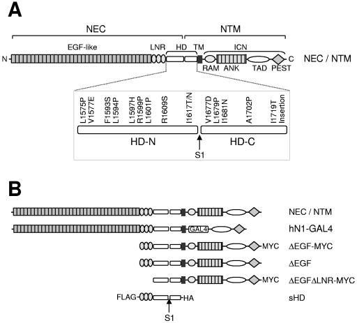
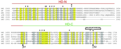
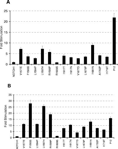
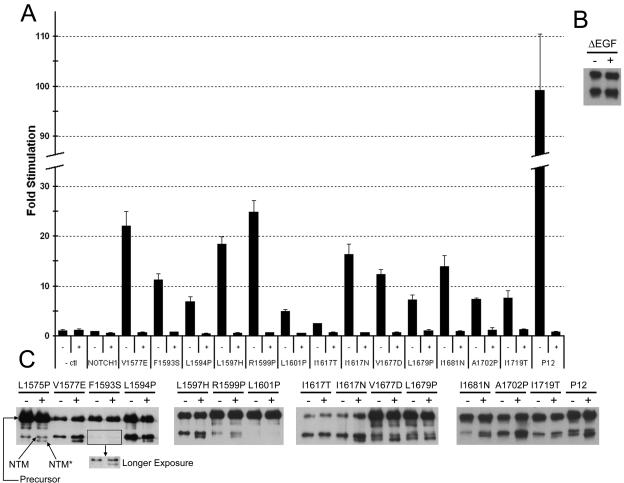
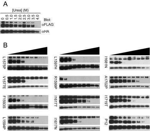
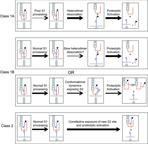
The NOTCH1 receptor is cleaved within its extracellular domain by furin during its maturation, yielding two subunits that are held together noncovalently by a juxtamembrane heterodimerization (HD) domain. Normal NOTCH1 signaling is initiated by the binding of ligand to the extracellular subunit, which renders the transmembrane subunit susceptible to two successive cleavages within and C terminal to the heterodimerization domain, catalyzed by metalloproteases and gamma-secretase, respectively. Because mutations in the heterodimerization domain of NOTCH1 occur frequently in human T-cell acute lymphoblastic leukemia (T-ALL), we assessed the effect of 16 putative tumor-associated mutations on Notch1 signaling and HD domain stability. We show here that 15 of the 16 mutations activate canonical NOTCH1 signaling. Increases in signaling occur in a ligand-independent fashion, require gamma-secretase activity, and correlate with an increased susceptibility to cleavage by metalloproteases. The activating mutations cause soluble NOTCH1 heterodimers to dissociate more readily, either under native conditions (n = 3) or in the presence of urea (n = 11). One mutation, an insertion of 14 residues immediately N terminal to the metalloprotease cleavage site, increases metalloprotease sensitivity more than all others, despite a negligible effect on heterodimer stability by comparison, suggesting that the insertion may expose the S2 site by repositioning it relative to protective NOTCH1 ectodomain residues. Together, these studies show that leukemia-associated HD domain mutations render NOTCH1 sensitive to ligand-independent proteolytic activation through two distinct mechanisms.


Figures

FIG. 1.
FIG. 1.
NOTCH1 expression constructs. (A) Schematic representation of the human NOTCH1 receptor. NEC, NOTCH1 extracellular subunit; NTM, NOTCH1 transmembrane subunit; LNR, domain comprising the three LIN12/Notch repeats; HD, heterodimerization domain; HD-N, N-terminal region of the HD domain; HD-C, C-terminal region of the HD domain; TM, transmembrane segment; ICN, intracellular NOTCH1; RAM, RAM domain; ANK, ankyrin repeat domain; TAD, transactivation domain; PEST, PEST domain; S1, furin cleavage site. The 16 leukemia-derived HD domain mutations described in this article are shown. (B) NOTCH1 polypeptides used in these studies.
FIG. 2.
FIG. 2.
Sequence alignment of the HD domains of various NOTCH receptors. Identical and conserved residues among the sequences are enclosed in yellow and gray boxes, respectively. h, human; x, Xenopus; m, mouse; and zf, zebrafish. Furin (S1) and metalloprotease (S2) cleavage sites are indicated below the sequence. Analyzed leukemia-derived mutations are identified above the sequence.
FIG. 3.
FIG. 3.
HD domain mutations result in activation of NOTCH1 signaling. HD domain mutations were introduced into (A) full-length or (B) ΔEGF NOTCH1 constructs. Luciferase assays were performed on U2OS cell lysates prepared from cells transfected in triplicate with 10 ng of empty pcDNA3 plasmid or plasmids encoding the indicated forms of (A) full-length or (B) ΔEGF NOTCH1, along with a luciferase reporter plasmid containing iterated CSL-binding sites, and an internal control plasmid expressing Renilla luciferase. Firefly luciferase activity from cell lysates was measured in triplicate, normalized, and expressed relative to the activity in extracts prepared from cells transfected with empty vector, which is arbitrarily set to a value of 1. The mutations L1575P and L1601P, which were previously shown to stimulate signaling in the context of full-length NOTCH1 (48), also stimulated signaling in the context of ΔEGF (data not shown).
FIG. 4.
FIG. 4.
HD domain mutations cause increased S2 and S3 cleavage. (A) Gamma secretase inhibitor (GSI) treatment abrogates the stimulatory effects of HD domain mutations in full-length NOTCH1. Immediately following transfection of U2OS cells with NOTCH1 expression plasmids encoding the indicated mutations, cells were treated with 1 μM compound E (GSI) or carrier alone. Trancriptional activation assays were performed 44 to 48 h after transfection, as described in the legend to Fig. 3. The increased signaling produced by the L1575P mutation was also inhibited by GSI in other experiments (data not shown). (B) U2OS cells transfected with wild-type ΔEGF-MYC NOTCH polypeptides were incubated in the presence of 1 μM compound E (GSI; +) or carrier (−) for 18 h prior to harvesting. NTM and NTM* polypeptides were then recovered by preparing immunoprecipitates from cell lysates using anti-MYC-coupled beads, resolved by SDS-PAGE under reducing conditions, and detected by Western blotting using an antibody against intracellular NOTCH1. (C) HD domain mutations were introduced into ΔEGF-MYC polypeptides, and the same assay was performed. Precursor, uncleaved NOTCH1 polypeptide; NTM, transmembrane subunit created by furin cleavage at site S1; and NTM*, S2 cleavage product.
FIG. 5.
FIG. 5.
Effect of HD domain mutations on subunit dissociation under native conditions. Conditioned media from 293T cells expressing secreted unmutated and mutated LNR-HD polypeptides (sHDs) were immunoprecipitated (IP) with anti-FLAG (α-FLAG) beads (top two panels) and analyzed following SDS-PAGE by Western blot analysis with the indicated antibodies. To better gauge whether the coprecipitation of the sHD subunits was affected by the mutations, a range of coprecipitated unmutated sHD subunits were run as a control. In addition to the sHD subunits, variable amounts of unprocessed sHD were detected in most of the conditioned media, including the normal control. Mutations inducing subunit dissociation of the HA-tagged HD-C subunit are denoted by stars. Comparable results were observed in parallel experiments in which immunoprecipitates were prepared with anti-HA (α-HA) beads (bottom pair of panels), and coprecipitation of the FLAG-tagged HD-N subunit was assessed by Western blotting.
FIG. 6.
FIG. 6.
Effect of HD domain mutations on subunit dissociation in the presence of urea. Conditioned media from 293T cells expressing (A) unmutated secreted NOTCH1 heterodimers or (B) mutated sHDs were immunoprecipitated with anti-HA (α-HA)-coupled beads, followed by 30 min of incubation at room temperature in buffer with different concentrations of urea (0 to 4 M). Subunit dissociation was evaluated by SDS-PAGE followed by Western blot analysis with antibodies specific for each of the two subunits. α-FLAG, anti-FLAG.
FIG. 7.
FIG. 7.
Proposed classification of leukemia-associated heterodimerization domain mutations. Activation by class 1 mutations occurs because of heterodimer dissociation or reduced heterodimer stability. Class 1A mutations cause overt heterodimer dissociation, while class 1B mutations cause either slow heterodimer dissociation or an increase in S2 site exposure by inducing local “breathing.” Class 2 mutations have no effect upon the stability of the heterodimer but result in proteolytic activation by positioning an exposed, mutation-associated S2 site away from protective residues that normally prevent metalloprotease access prior to ligand binding. The thickness of the arrow represents the estimated flux of molecules through the indicated step. HD-N, red; HD-C, blue; S1 site, filled triangle; S2 site, open triangle. The dashed line in the bottom panel represents the inserted sequence, and x indicates the position of the original S2 site ablated by the insertion.

References

    1. Ahimou, F., L. P. Mok, B. Bardot, and C. Wesley. 2004. The adhesion force of Notch with Delta and the rate of Notch signaling. J. Cell Biol. 167:1217-1229. - PMC - PubMed
    1. Anderson, G., J. Pongracz, S. Parnell, and E. J. Jenkinson. 2001. Notch ligand-bearing thymic epithelial cells initiate and sustain Notch signaling in thymocytes independently of T cell receptor signaling. Eur. J. Immunol. 31:3349-3354. - PubMed
    1. Artavanis-Tsakonas, S., M. D. Rand, and R. J. Lake. 1999. Notch signaling: cell fate control and signal integration in development. Science 284:770-776. - PubMed
    1. Aster, J. C., E. S. Robertson, R. P. Hasserjian, J. R. Turner, E. Kieff, and J. Sklar. 1997. Oncogenic forms of NOTCH1 lacking either the primary binding site for RBP-Jkappa or nuclear localization sequences retain the ability to associate with RBP-Jkappa and activate transcription. J. Biol. Chem. 272:11336-11343. - PubMed
    1. Aster, J. C., L. Xu, F. G. Karnell, V. Patriub, J. C. Pui, and W. S. Pear. 2000. Essential roles for ankyrin repeat and transactivation domains in induction of T-cell leukemia by Notch1. Mol. Cell. Biol. 20:7505-7515. - PMC - PubMed

Publication types

MeSH terms