. 2006 Jun;26(12):4664-74.
doi: 10.1128/MCB.02253-05.

Concerted functions of Gab1 and Shp2 in liver regeneration and hepatoprotection

Affiliations

Concerted functions of Gab1 and Shp2 in liver regeneration and hepatoprotection

Emilie A Bard-Chapeau et al. Mol Cell Biol. 2006 Jun.

Abstract

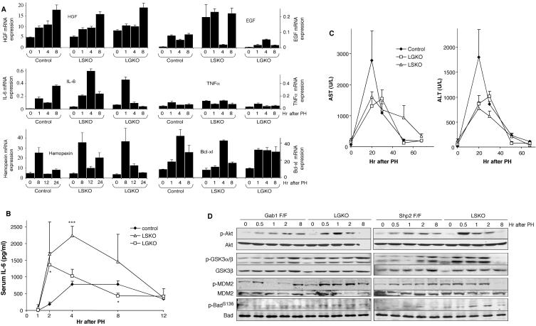
Liver regeneration is a rapid and concerted response to injury, in which growth factor-generated intracellular signals result in activation of transcription factors, DNA synthesis, and hepatocyte proliferation. However, the link between cytoplasmic signals resulting in proliferative response to liver injury remains to be elucidated. We show here that association of Gab1 adaptor protein and Shp2 tyrosine phosphatase is a critical event at the early phase of liver regeneration. Partial hepatectomy (PH) rapidly and transiently induced assembly of a complex comprising Shp2 and tyrosine-phosphorylated Gab1 in wild-type hepatocytes. Consistently, liver-specific Shp2 knockout (LSKO) and liver-specific Gab1 knockout (LGKO) mice displayed very similar phenotypes of defective liver regeneration triggered by PH, including blunted extracellular signal-regulated kinase 1/2 (Erk1/2) activation, decreased expression of immediate-early genes, and reduced levels of cyclins A, E, and B1, as well as suppression of hepatocyte proliferation. In contrast, the Akt and interleukin-6/Stat3 pathways were up-regulated posthepatectomy in LSKO and LGKO mice, accompanied by improved hepatoprotection. Collectively, this study establishes the physiological significance of the Gab1/Shp2 link in promoting mitogenic signaling through the Erk pathway in mammalian liver regeneration.


Figures

FIG. 1.
FIG. 1.
Selective deletion of Shp2 in the hepatocytes of LSKO mice. (A) Genotyping of mice by PCR. Genomic DNA was extracted from liver (L), spleen (S), white adipose tissue (Wat), gut (G), lung (Lu), pancreas (P), kidney (K), skeletal muscle (M), pituitary gland (Pi), and hypothalamus (H) of an LSKO mouse (Cre/+ Shp2flox/flox [Shp2 F/F]) and a Shp2flox/flox mouse (+/+ F/F). The Shp2flox allele produced a 3-kb fragment, and the Shp2 allele gave rise to a 1-kb fragment. Deletion of Shp2 exon 4 was detected only in the livers of LSKO mice. (B) (Top) Immunoblot analysis of Shp2 protein expression in liver, kidney, heart, white adipose tissue (wat), and skeletal muscle isolated from mice of different genotypes (wt, wild type). (Bottom) Immunoblotting of Shp2 in liver lysates was done with anti-Erk2 blot as a loading control. Shp2 protein content was dramatically reduced in the liver lysate of LSKO mice. (C) Immunoblot analysis of Shp2 in hepatocytes isolated from control or LSKO mice, with tubulin used as a loading control.
FIG. 2.
FIG. 2.
Reduced hepatocyte proliferation in the livers of LSKO and LGKO mice posthepatectomy. (A) Quantification of BrdU labeling. Control (Shp2flox/flox or Gab1flox/flox), LSKO, and LGKO mice were subjected to partial hepatectomy and were injected with BrdU 2 h before harvest. Animals were sacrificed at the indicated time points, and the number of BrdU-positive hepatocytes was quantified. Values for the LSKO or LGKO mice that were significantly different from the values for the control (Shp2flox/flox or Gab1flox/flox) mice by a two-tailed unpaired t test are indicated as follows: *, P < 0.05; **, P < 0.01; ***, P < 0.001. Values are expressed as means ± standard errors of the means (SEMs) (error bars) (n = 3 to 7). (B) Representative samples of BrdU immunolabeling (green) were taken at 40, 44, and 68 h after PH. DAPI-stained nuclei are shown in red. Magnification, ×100. (C) Recuperation of the liver mass after PH. Liver weight per body weight was measured at 24, 48, and 72 h after surgery. Values are expressed as means ± SEMs (error bars) (n = 3 to 7). (D) Immunoblot analysis of cyclins A, B1, E, and D (with Erk2 as a loading control) was performed with liver lysates collected at the indicated time points after PH. Signals were quantified using ImageQuant software, with the strongest signal set at the value of 1.
FIG. 3.
FIG. 3.
Association of Gab1 and Shp2 at the plasma membrane after PH. (A) Control mice (Gab1flox/flox [Gab1 F/F]) were subjected to partial hepatectomy, and remnant livers were harvested at the indicated time points after surgery. Liver extracts were immunoprecipitated with an anti-Gab1 antibody (IP Gab1) and immunoblotted with antibodies against phosphotyrosine (PY), Gab1, or Shp2. (B) Liver extracts from Gab1flox/flox (Gab1 F/F) and LGKO mice and Shp2flox/flox (Shp2 F/F) and LSKO mice were immunoprecipitated with the Gab1 antibody and immunoblotted with antibodies to PY or Gab1. (C) Liver extracts were immunoprecipitated with the Gab1 antibody and immunoblotted with antibodies to the p85 subunit of PI3K, Grb2, PLC-γ, or Gab1. (D). RasGAP was precipitated with its specific antibody, and the immunoprecipitates were immunoblotted with antibodies to PY or RasGAP. Signals were quantified using ImageQuant software, with the strongest signal of controls set at the value of 1. (E) Cytoplasmic and plasma membrane-enriched fractions were isolated from livers collected at 0 and 0.5 h after PH. The lysates were subjected to immunoblotting with antibodies against Gab1, Shp2, RasGAP, and Grb2, with Met and Erk2 used as loading controls.
FIG. 4.
FIG. 4.
Impaired activation of Erk. (A) Liver extracts from Shp2flox/flox (Shp2 F/F) and LSKO mice and Gab1flox/flox (Gab1 F/F) and LGKO mice were prepared from 0 to 8 h after PH and immunoblotted with antibodies against p-Erk1/2, Erk2, p-p38-MAPK (p-p38), p38-MAPK (p38), p-Jnk1/2, and Jnk1/2. (B) Graphic representation of p-Erk2 levels, normalized against Erk2 protein amounts (n = 2 for each control; n = 2 for each knockout). (C) Immunoblot analysis of p-Erk1/2 and Erk2 was performed for liver extracts prepared after portal vein injection of saline (−) or IL-6, EGF, or HGF (+) into control and mutant mice. (D) Liver extracts were prepared from 0 and 8 h after PH and immunoblotted with antibodies against p-Met, Met, p-EGFR, and EGFR.
FIG. 5.
FIG. 5.
Impaired expression of immediate-early genes. (A) Liver extracts from Shp2flox/flox (Shp2 F/F) and LSKO mice and Gab1flox/flox (Gab1 F/F) and LGKO mice were prepared and immunoblotted with an antibody to pan-Fos. The migration positions of the different family members range from 55 to 62 kDa. (B) Expression levels of c-Fos, c-Jun, c-Myc, and C/EBPβ mRNAs. Total RNAs were isolated from hepatectomized livers and assessed by real-time RT-PCR. Values are expressed as means ± standard errors of the means (SEMs) (error bars). The results shown are from three different mice per time point and genotype.
FIG. 6.
FIG. 6.
Enhanced Stat3 activation. (A) Stat3 mRNA levels from control (Shp2flox/flox or Gab1flox/flox), LSKO, and LGKO mice. Real-time RT-PCR analysis was performed as described in the legend to Fig. 5B. (B) Induction of p-Tyr705Stat3 was measured at 0 to 8 h after PH by immunoblotting of liver extracts. The liver extracts were from Shp2flox/flox (Shp2 F/F) and LSKO mice and Gab1flox/flox (Gab1 F/F) and LGKO mice. (C) Graphic representation of p-Tyr705Stat3 levels normalized against Stat3 protein amounts (n = 2 for each control; n = 2 for each knockout). (D) Stat3 DNA binding activity was assessed by gel mobility shift assay with nuclear extracts. The leftmost lane shows a supershift (ss) when Stat3 antibody was added to control extract prepared at 5 h. The next lane shows a competition (Comp.) assay in which 50 ng of cold probe was added to the control extract at 5 h.
FIG. 7.
FIG. 7.
Enhanced IL-6 production and p-Akt levels. (A) Levels of HGF, EGF, IL-6, TNF-α, Stat3, hemopexin, and Bcl-xl mRNAs were assessed by real-time RT-PCR. Values are expressed as means ± standard errors of the means (SEMs) (error bars). The results are representative of three mice. (B) Circulating blood levels of IL-6 were quantified at the indicated time points posthepatectomy. Values for the LSKO or LGKO mice that were significantly different from the values for the control (Shp2flox/flox or Gab1flox/flox) mice by a two-tailed unpaired t test are indicated as follows: *, P < 0.05; ***, P < 0.001. Values are expressed as means ± SEMs (error bars) (n = 3 to 5). (C) Circulating blood levels of AST and ALT (in units per liter) were quantified at different time points after surgery. Values are expressed as means ± SEMs (error bars) (n = 3 or 4). (D) p-Akt, p-GSK3, p-MDM2, and p-Bad levels were evaluated at 0 to 8 h after PH by immunoblotting of liver extracts from Gab1flox/flox (Gab1 F/F) and LGKO mice and Shp2flox/flox (Shp2 F/F) and LSKO mice. Akt, GSK3, MDM2, and Bad were used as loading controls.

References

    1. Aldeguer, X., F. Debonera, A. Shaked, A. M. Krasinkas, A. E. Gelman, X. Que, G. A. Zamir, S. Hiroyasu, K. K. Kovalovich, R. Taub, and K. M. Olthoff. 2002. Interleukin-6 from intrahepatic cells of bone marrow origin is required for normal murine liver regeneration. Hepatology 35:40-48. - PubMed
    1. Bard-Chapeau, E. A., A. L. Hevener, S. Long, E. E. Zhang, J. M. Olefsky, and G. S. Feng. 2005. Deletion of Gab1 in the liver leads to enhanced glucose tolerance and improved hepatic insulin action. Nat. Med. 11:567-571. - PubMed
    1. Bardin, A. J., and A. Amon. 2001. Men and sin: what's the difference? Nat. Rev. Mol. Cell Biol. 2:815-826. - PubMed
    1. Bladt, F., D. Riethmacher, S. Isenmann, A. Aguzzi, and C. Birchmeier. 1995. Essential role for the c-met receptor in the migration of myogenic precursor cells into the limb bud. Nature 376:768-771. - PubMed
    1. Blindenbacher, A., X. Wang, I. Langer, R. Savino, L. Terracciano, and M. H. Heim. 2003. Interleukin 6 is important for survival after partial hepatectomy in mice. Hepatology 38:674-682. - PubMed

Publication types

MeSH terms

LinkOut - more resources