. 2006 Jun;26(12):4729-45.
doi: 10.1128/MCB.01973-05.

Slm1 and slm2 are novel substrates of the calcineurin phosphatase required for heat stress-induced endocytosis of the yeast uracil permease

Affiliations

Slm1 and slm2 are novel substrates of the calcineurin phosphatase required for heat stress-induced endocytosis of the yeast uracil permease

Geert Bultynck et al. Mol Cell Biol. 2006 Jun.

Abstract

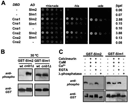
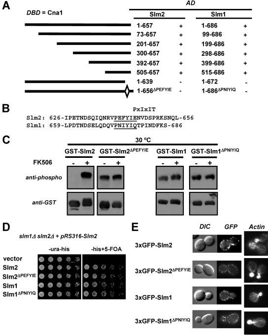
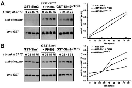
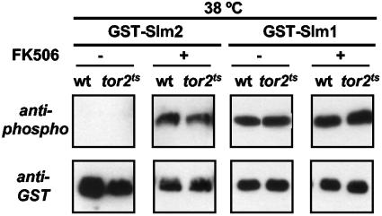
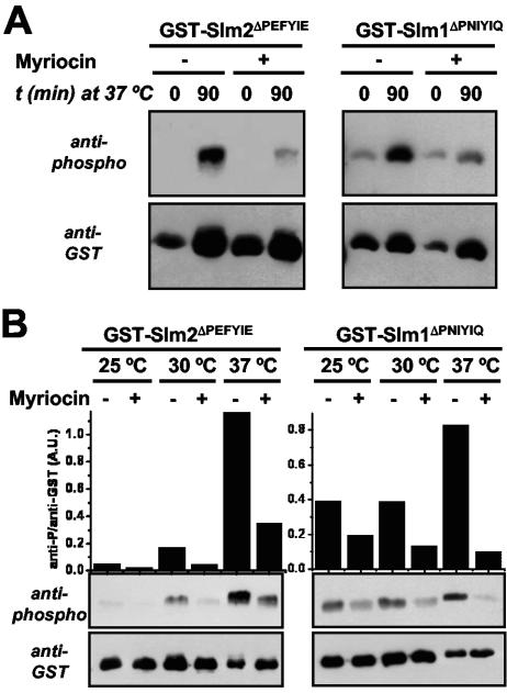
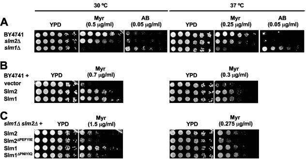
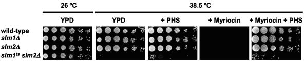
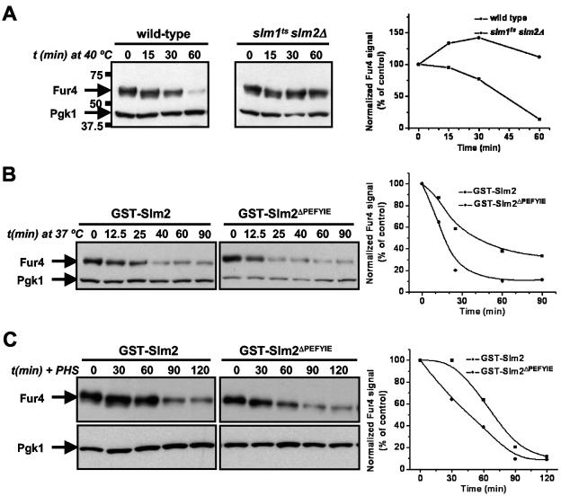
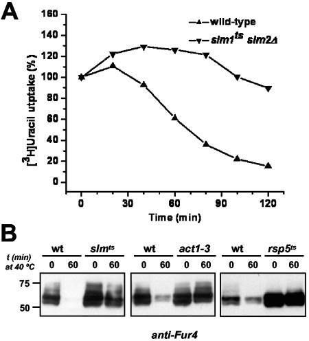
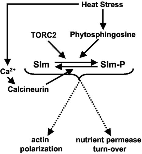
The Ca2+/calmodulin-dependent phosphatase calcineurin promotes yeast survival during environmental stress. We identified Slm1 and Slm2 as calcineurin substrates required for sphingolipid-dependent processes. Slm1 and Slm2 bind to calcineurin via docking sites that are required for their dephosphorylation by calcineurin and are related to the PXIXIT motif identified in NFAT. In vivo, calcineurin mediates prolonged dephosphorylation of Slm1 and Slm2 during heat stress, and this response can be mimicked by exogenous addition of the sphingoid base phytosphingosine. Slm proteins also promote the growth of yeast cells in the presence of myriocin, an inhibitor of sphingolipid biosynthesis, and regulation of Slm proteins by calcineurin is required for their full activity under these conditions. During heat stress, sphingolipids signal turnover of the uracil permease, Fur4. In cells lacking Slm protein activity, stress-induced endocytosis of Fur4 is blocked, and Fur4 accumulates at the cell surface in a ubiquitinated form. Furthermore, cells expressing a version of Slm2 that cannot be dephosphorylated by calcineurin display an increased rate of Fur4 turnover during heat stress. Thus, calcineurin may modulate sphingolipid-dependent events through regulation of Slm1 and Slm2. These findings, in combination with previous work identifying Slm1 and Slm2 as targets of Mss4/phosphatidylinositol 4,5-bisphosphate and TORC2 signaling, suggest that Slm proteins integrate information from a variety of signaling pathways to coordinate the cellular response to heat stress.


Figures

FIG. 1.
FIG. 1.
Slm1 and Slm2 interact with and are dephosphorylated by calcineurin. (A) Slm1 and Slm2 interact with Cna1 and Cna2 in yeast two-hybrid assays. A strain (PJ69-4A) containing GAL1-HIS3, GAL2-ADE2, and GAL7-lacZ reporter genes was transformed with combinations of GAL4 DNA-binding domain fusions of CNA1 and CNA2 (DBD) and GAL4 activation domain fusions of SLM1 and SLM2 (AD), as indicated. Serial dilutions of saturated cultures were plated on synthetic medium containing histidine and adenine (+his+ade) or lacking histidine (-his) or adenine (-ade), and incubated for 3 days at 30°C. β-Galactosidase (βgal) activity in the different strains was measured in liquid cultures as described in Materials and Methods and reported as units/μg of protein. (B) Slm1 and Slm2 are both dephosphorylated in vivo by calcineurin. Wild-type cells (wt; YPH499) and cells lacking the B subunit of calcineurin (cnb1Δ; DD12) expressing GST-SLM2 (GBY030 and GBY032) or GST-SLM1 (GBY031 and GBY033) were grown to log phase. GST fusion proteins were purified from extracts, subjected to SDS-PAGE, and Western blotted with anti-phospho or anti-GST antibody. (C) Slm1 and Slm2 are both dephosphorylated in vitro by calcineurin. GST-Slm1 (GBY032) and GST-Slm2 (GBY033) purified from cnb1Δ cells were treated with recombinant calcineurin, calmodulin (CaM), CaCl2, EGTA, or λ-phosphatase as indicated in the left-hand lanes (see Materials and Methods). These samples, together with untreated GST-Slm1 and GST-Slm2 purified from cnb1Δ cells, were then subjected to SDS-PAGE and Western blotted with anti-phospho and anti-GST antibodies.
FIG. 2.
FIG. 2.
Calcineurin interacts with Slm1 and Slm2 via a C-terminal PXIXIT-related motif. (A) Mapping of the calcineurin-docking site on Slm1 and Slm2 via yeast two-hybrid assays. The PJ69-4A strain containing the GAL4 DNA-binding domain fusion of CNA1 (DBD) was transformed with different truncations or internal deletions of SLM1 and SLM2. Positive interactions were identified as growth on synthetic medium lacking histidine (3 days at 30°C). GAL4 activation domain fusions (AD) that interacted with Cna1 are indicated as “+”, and those that did not interact are indicated as “−.” (B) Relevant parts of the amino acid sequences of Slm2 and Slm1 with the conserved calcineurin-docking sites, PXIXIT-related motifs, underlined. (C) Deletion of the calcineurin-docking site abolishes the calcineurin-dependent dephosphorylation of Slm1 and Slm2. Wild-type GST-SLM2 (GBY115) and GST-SLM1 (GBY117) and their counterparts lacking the calcineurin-docking site, GST-SLM2ΔPEFYIE (GBY116) and GST-SLM1ΔPNIYIQ (GBY118), were expressed in a strain deleted for slm1Δ and slm2Δ. Cells were grown in selective synthetic medium supplemented with 4% raffinose. One culture was treated with FK506 (as indicated). GST fusion proteins were purified from cell extracts, subjected to SDS-PAGE, and Western blotted with anti-phospho and anti-GST antibodies. (D) Deletion of the calcineurin-docking site in SLM1 or SLM2 does not impair growth. An slm1Δ slm2Δ strain expressing SLM2 under the control of its own promoter on a pRS316 plasmid (GBY059) was transformed with either pRS313 (vector; GBY152), pRS313-SLM2 (Slm2; GBY153), pRS313-SLM2ΔPEFYIE (Slm2 ΔPEFYIE; GBY154), pRS313-SLM1 (Slm1; GBY155), or pRS313-SLM1ΔPNIYIQ (Slm1ΔPNIYIQ; GBY156). Serial dilutions of saturated cell cultures were plated on synthetic medium lacking uracil and histidine (-ura-his) and on synthetic medium lacking histidine but supplemented with 5FOA (-his+5-FOA). Cells were grown for 3 days at 30°C. (E) Localization of Slm proteins and actin cytoskeleton stabilization by Slm proteins are not dependent on the calcineurin-docking site. slm1Δ slm2Δ cells expressing GFP-SLM2 (GBY104), GFP-SLM2ΔPEFYIE (GBY105), GFP-SLM1 (GBY106), or GFP-SLM1ΔPNIYIQ (GBY107) were grown to log phase and visualized by differential interference microscopy (DIC) and by fluorescence microscopy (GFP). Actin cytoskeleton was visualized with Texas Red-X phalloidin in formaldehyde-fixed cells.
FIG. 3.
FIG. 3.
Phosphorylation of Slm1 and Slm2 is increased during heat stress when their calcineurin-dependent dephosphorylation is compromised. (A) GST-SLM2 (GBY115) and GST-SLM2ΔPEFYIE (GBY116) were expressed in a strain lacking slm1 and slm2. Cells were grown at 25°C in selective synthetic medium and expression was induced with 2% galactose. One culture was treated with FK506 (2 μg/ml) as indicated. Cells were shifted to 37°C and samples were taken at indicated time points (t). GST fusion proteins were purified from cell extracts and subjected to SDS-PAGE and Western blotting with anti-phospho and anti-GST antibodies. Immunoreactive bands were quantified using ImageJ software. The ratio of the anti-phospho signal over the anti-GST signal was plotted in arbitrary units (A.U.) as a function of time. (B) An approach similar to that described for panel A was taken for GST-SLM1 (GBY117) and GST-SLM1ΔPNIYIQ (GBY118).
FIG. 4.
FIG. 4.
Phytosphingosine mimics the heat stress-induced phosphorylation of Slm1 and Slm2. GST-SLM2 (GBY115), GST-SLM2ΔPEFYIE (GBY116), GST-SLM1 (GBY117), and GST-SLM1ΔPNIYIQ (GBY118) were expressed in a strain lacking slm1 and slm2. Cells were grown at 25°C in selective synthetic medium and expression was induced with 2% galactose. Samples were taken just before the addition of 20 μM phytosphingosine (0 min) and at time points (t) 45, 90, and 120 min after the addition of phytosphingosine. GST fusion proteins were purified from cell extracts and subjected to SDS-PAGE and Western blotting with anti-phospho and anti-GST antibodies. Immunoreactive bands were quantified as described in Materials and Methods. The ratio of the anti-phospho signal over the anti-GST signal was plotted in arbitrary units (A.U.).
FIG. 5.
FIG. 5.
The heat stress-induced increase in Slm1 and Slm2 phosphorylation is independent of Tor2p. Overnight cultures of wild-type (wt; JK9-3da) and tor2ts (SH121) cells expressing either GST-Slm2 or GST-Slm1 were diluted to an OD600 of 0.2 and grown at 25°C for 4 h. To one set of cultures, FK506 (final concentration, 2 μg/ml) or solvent (90% ethanol/10% Tween 20) was added and cells were shifted to 38°C. After 30 min, galactose (2%) was added to induce protein expression and cells were grown for another 4 h at 38°C. GST fusion proteins were purified from cell extracts and subjected to SDS-PAGE and Western blotting with anti-phospho and anti-GST antibodies.
FIG. 6.
FIG. 6.
Myriocin inhibits phosphorylation of Slm1 and Slm2 during heat stress and steady-state growth conditions. GST-SLM2ΔPEFYIE (GBY116) and GST-SLM1ΔPNIYIQ (GBY118) were expressed in an slm1Δ slm2Δ strain. (A) Cells were grown at 25°C in selective synthetic medium for 4 h and protein expression was induced with 2% galactose. Thirty minutes before the shift from 25°C to 37°C, myriocin (2 μg/ml) was added to one set of cultures and solvent (methanol) to the other. Samples of the different cultures were taken at time points (t) just before the temperature shift and 90 min after the shift in temperature. GST fusion proteins were purified from cell extracts and subjected to SDS-PAGE and Western blotting with either an anti-phospho or an anti-GST antibody. (B) Cells were grown at either 25°C, 30°C, or 37°C in selective synthetic medium. Myriocin (2 μg/ml) was added 30 min before the cells were incubated with 2% galactose for 4 h. GST fusion proteins were purified from cell extracts and analyzed via Western immunoblotting with either an anti-phospho or an anti-GST antibody. Immunoreactive bands were quantified as described in Materials and Methods. A.U., arbitrary units.
FIG. 7.
FIG. 7.
Slm1 and Slm2 modulate growth of yeast in the presence of drugs that perturb sphingolipid biosynthesis. (A) Saturated cultures of wild-type cells (BY4741) and cells lacking either slm1 (slm1Δ; VHY66) or slm2 (slm2Δ; VHY61) were serially diluted and spotted onto YPD, YPD plus myriocin (Myr), or YPD plus aureobasidin A (AB) and grown for 2 to 4 days at either 30°C or 37°C. (B) Wild-type cells containing either pRS313 (vector; GBY160) or pRS313 expressing Slm2 (GBY161) and Slm1 (GBY162) under the control of their own promoters were grown to saturation in selective synthetic medium. Saturated cultures were serially diluted and were spotted onto YPD with or without myriocin. Plates were incubated at either 30°C or 37°C for 2.5 days. (C) slm1Δ slm2Δ cells expressing SLM2 (GBY100), SLM2ΔPEFYIE (GBY101), SLM1 (GBY102), or SLM1ΔPNIYIQ (GBY103) under the control of their own promoters from pRS313 were grown to saturation in selective synthetic medium. Saturated cultures were serially diluted and were spotted onto YPD or YPD supplemented with 1.5 μg/ml myriocin. Cells were grown for 3 to 4 days at either 30°C or 37°C as indicated.
FIG. 8.
FIG. 8.
Slm proteins are downstream of phytosphingosine. Saturated cultures of wild-type (SEY6210), slm1Δ (AAY1602), slm2Δ (AAY1610), and slm1ts slm2Δ (AAY1623.2) cells were serially diluted and spotted onto YPD, YPD plus 20 μM phytosphingosine (PHS), YPD plus 0.4 μg/ml myriocin, or YPD plus 0.4 μg/ml myriocin plus 20 μM phytosphingosine and grown for 2.5 days at the indicated temperatures. All plates contained 0.05% NP-40, which was used for obtaining even distributions of phytosphingosine in the plates.
FIG. 9.
FIG. 9.
Slm function is required for heat shock-induced Fur4 turnover. (A) Wild-type cells (SEY6210.1) and slm1ts slm2Δ cells (AAY1623.2) carrying pVTu-FUR4 (GBY179 and GBY180, respectively) were shifted from 25°C to 40°C and samples were taken at time points (t) just prior to (0 min) and 15, 30, and 60 min after heat shock. Cell lysates were analyzed by SDS-PAGE and immunoblotting with anti-Fur4 and anti-PGK antibodies. Pgk1 was used as a loading control, which also shows the absence of nonspecific protein degradation by heat shock. The numbers to the left of the gels indicate the molecular masses of the protein standards. Immunoreactive bands were quantified as described in Materials and Methods. The ratio of the anti-Fur4 signal to the anti-Pgk1p signal is shown as a function of time. The value at 0 min was used as the reference value and set at 100%. (B) slm1Δ slm2Δ cells expressing either GST-SLM2 or GST-SLM2ΔPEFYIE and overexpressing FUR4 (GBY115 and GBY116, respectively) were shifted from 25°C to 40°C and sampled at the indicated time points. Cell lysates were analyzed by immunoblotting and quantified as described for panel A. (C) GBY115 and GBY116 were grown at 30°C, incubated with 20 μM phytosphingosine, and sampled at the indicated time points. Lysates were analyzed and signals quantified as described for panel A.
FIG. 10.
FIG. 10.
Fur4 is stabilized at the plasma membrane in the ubiquitinated form in cells lacking Slm protein activity. Wild-type cells (SEY6210.1) and slm1ts slm2Δ cells (AAY1623.2) carrying pVTu-FUR4 (GBY179 and GBY180, respectively) were shifted to 40°C for 10 min before the addition of cycloheximide (100 μg/ml). (A) Uracil uptake was measured at the times indicated. Results are percentages of initial activities. (B) Plasma membrane fractions were prepared from cycloheximide-treated cells before and 60 min after a shift from 25°C to 40°C and analyzed for uracil permease (anti-Fur4) by Western immunoblotting. Strains are as follows: left panel, wild type (wt) (SEY6210.1) and slmts (AAY1623.2); middle panel, wild type (NY13) and act1-3 (NY279); right panel, wild type (FY56) and rsp5ts (FW1808).
FIG. 11.
FIG. 11.
Regulation of actin cytoskeleton organization and nutrient permease turnover by Slm protein functioning in response to heat stress. See the text for a discussion of the model.

References

    1. Aramburu, J., F. Garcia-Cozar, A. Raghavan, H. Okamura, A. Rao, and P. G. Hogan. 1998. Selective inhibition of NFAT activation by a peptide spanning the calcineurin targeting site of NFAT. Mol. Cell 1:627-637. - PubMed
    1. Aramburu, J., A. Rao, and C. B. Klee. 2000. Calcineurin: from structure to function. Curr. Top. Cell. Regul. 36:237-295. - PubMed
    1. Aramburu, J., M. B. Yaffe, C. Lopez-Rodriguez, L. C. Cantley, P. G. Hogan, and A. Rao. 1999. Affinity-driven peptide selection of an NFAT inhibitor more selective than cyclosporin A. Science 285:2129-2133. - PubMed
    1. Audhya, A., R. Loewith, A. B. Parsons, L. Gao, M. Tabuchi, H. Zhou, C. Boone, M. N. Hall, and S. D. Emr. 2004. Genome-wide lethality screen identifies new PI4,5P(2) effectors that regulate the actin cytoskeleton. EMBO J. 23:3747-3757. - PMC - PubMed
    1. Ausubel, F. M, R. Brent, R. E. Kingston, D. D. Moore, J. G. Seidman, J. A. Smith, and K. Struhl. 1987. Current protocols in molecular biology. John Wiley & Sons, New York, N.Y.

Publication types

MeSH terms