. 2006 Jun;26(12):4746-57.
doi: 10.1128/MCB.00959-05.

Repression of the antiapoptotic molecule galectin-3 by homeodomain-interacting protein kinase 2-activated p53 is required for p53-induced apoptosis

Affiliations

Repression of the antiapoptotic molecule galectin-3 by homeodomain-interacting protein kinase 2-activated p53 is required for p53-induced apoptosis

Barbara Cecchinelli et al. Mol Cell Biol. 2006 Jun.

Abstract

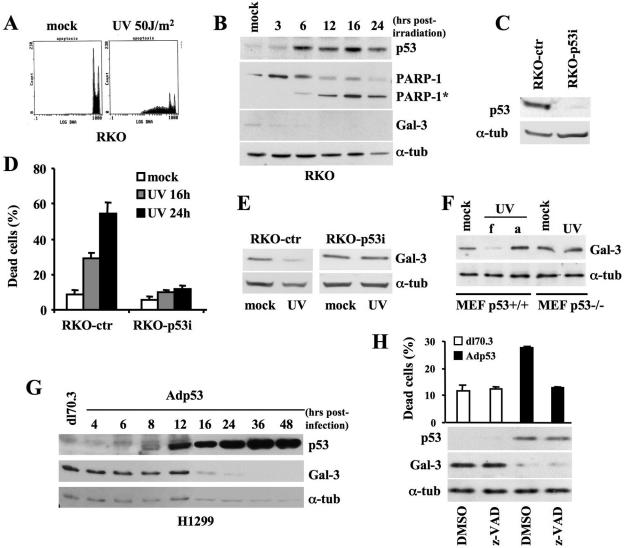
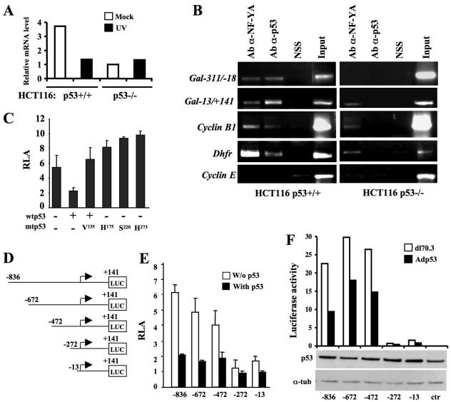
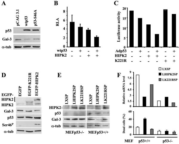
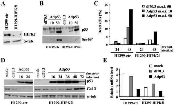
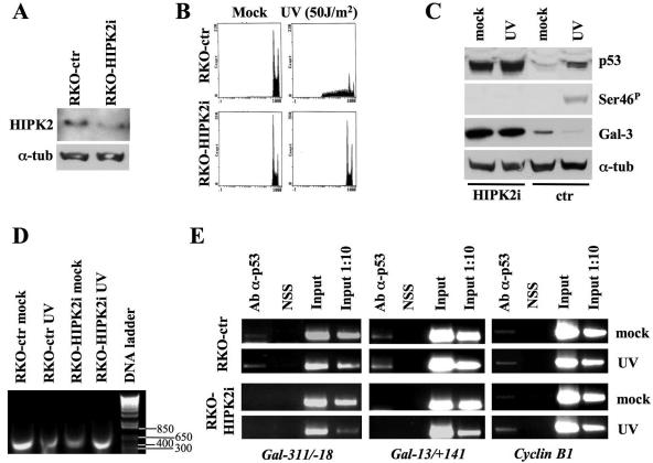
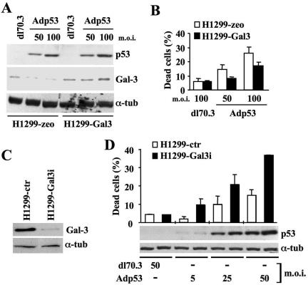
Galectin 3 (Gal-3), a member of the beta-galactoside binding lectin family, exhibits antiapoptotic functions, and its aberrant expression is involved in various aspects of tumor progression. Here we show that p53-induced apoptosis is associated with transcriptional repression of Gal-3. Previously, it has been reported that phosphorylation of p53 at Ser46 is important for transcription of proapoptotic genes and induction of apoptosis and that homeodomain-interacting protein kinase 2 (HIPK2) is specifically involved in these functions. We show that HIPK2 cooperates with p53 in Gal-3 repression and that this cooperation requires HIPK2 kinase activity. Gene-specific RNA interference demonstrates that HIPK2 is essential for repression of Gal-3 upon induction of p53-dependent apoptosis. Furthermore, expression of a nonrepressible Gal-3 prevents HIPK2- and p53-induced apoptosis. These results reveal a new apoptotic pathway induced by HIPK2-activated p53 and requiring repression of the antiapoptotic factor Gal-3.


Figures

FIG. 1.
FIG. 1.
Gal-3 expression is repressed in p53-mediated apoptosis. (A) RKO cells were exposed to UV (50 J/m2) and collected 24 h later for cell cycle analysis upon PI staining. DNA-content analysis by flow cytometry of one indicative experiment is reported. (B) Immunoblotting kinetics of p53, PARP-1 and its cleaved form (PARP-1*), and Gal-3 at the indicated times after UV irradiation (50 J/m2). α-Tubulin expression is used as a loading control. (C) Immunoblots with antibody to p53 on RKO cells stably transfected with pSUPER-ctr and pSUPER-p53 vectors. α-Tubulin was used as a loading control. (D) Percentage of dead cells determined by trypan blue exclusion in the indicated cells at the indicated times after UV irradiation. Means ± standard deviations of three independent experiments are reported. (E) Immunoblot of Gal-3 on the indicated cells after UV irradiation. α-Tubulin was used as a loading control. (F) Immunoblot of Gal-3 48 h after UV irradiation of MEF from p53+/+ and p53−/− mice. Due to the small amount of apoptosis in these primary cells, the UV-irradiated MEFs were harvested, with the dead, floating cells (f) kept separate from the adherent ones (a). In the case of p53−/− MEFs, only adherent cells were present. (G) Immunoblotting kinetics of p53 and Gal-3 after infection of p53-null H1299 cells with Adp53 recombinant adenovirus or dl70.3 control virus at an MOI of 50. α-Tubulin was used as a loading control. (H) H1299 cells were infected as described in panel G in the presence of the pan-caspase inhibitor z-VAD or its solvent. Percentage of dead cells was calculated as described for panel D at 24 h postinfection when cells were harvested for immunoblot analyses, as described for panel G. α-tub, α-tubulin.
FIG. 2.
FIG. 2.
p53 represses Gal-3 at the transcriptional level. (A) Analysis of Gal-3 mRNA expression. UV irradiation was performed as described in the legend of Fig. 1A, and RNA was collected from mock-infected cells and at 16 h postirradiation. Real-time RT-PCR was performed using the 18S RNA as a control. (B) Cross-linked chromatin from the indicated cells was immunoprecipitated with sheep antibody to p53 (Ab-α-p53), normal sheep serum, or rabbit antibody to NF-YA (Ab-α-NF-YA) and analyzed by PCR with primers specific for the indicated promoters. Input corresponds to nonimmunoprecipitated cross-linked chromatin. (C) LGALS3 promoter activity. Coexpression studies were performed in human H1299 cells using the p(−836/+141)LGALS3-Luc reporter construct, together with wtp53, dnp53, or the indicated mutant p53. Means ± standard deviations of at least seven independent experiments are reported. (D) Schematic representation of the human LGALS3 promoter fragments cloned upstream of the firefly luciferase reporter gene in the pGL-3-Basic vector. (E) Luciferase activity of the reporter vectors shown in panel D and indicated with the most-5′ base of the LGALS3 promoter sequence cloned into the relative vectors, in the presence or absence of wtp53. Means ± standard deviations of at least seven independent experiments are reported. (F) Luciferase activity of the same reporter vectors stably transfected into H1299 cells. Each mixed population, indicated as in panel E was infected at an MOI of 50 with dl70.3 control or Adp53 virus. The expression of exogenous p53 in each mixed population is shown by the immunoblot. One indicative experiment out of three performed is reported. α-tub, α-tubulin; NSS, normal sheep serum.
FIG. 3.
FIG. 3.
p53 phosphorylation at Ser46 by HIPK2 is required for p53-mediated repression of Gal-3. (A) Immunoblots with antibodies to p53 and Gal-3 on H1299 cells transfected with pCAG3.1 control, pCAG3.1-wtp53, or pCAG3.1-p53S46A mutant vector. α-Tubulin was used as a loading control. (B) Coexpression studies were performed in human H1299 cells by transfecting the p(−836/+141)LGALS3-Luc reporter vector with wtp53 and/or HIPK2 expression vectors. Means ± standard deviations of at least seven independent experiments are reported. (C) Luciferase activity of the same p(−836/+141)LGALS3-Luc reporter vector stably transfected into H1299 cells. The polyclonal population was infected with Adp53 at MOI of 10 and transiently transfected with HIPK2- or K221R mutant-expressing vectors. The results of one indicative experiment out of three performed are reported. (D) Immunoblot of total p53 and p53 phosphorylation at Ser46 (Ser-46P), HIPK2, and Gal-3 in p53-positive HEK293 cells transiently transfected with pEGFP vectors encoding EGFP-tagged wild-type HIPK2 or its KD K221R mutant. α-Tubulin was used as a loading control. (E and F) MEFs from p53+/+ and p53−/− mice were infected with recombinant LHIPK2SP, LK221RSP, or control LXSP retroviruses. At 36 h postinfection, cells were harvested to perform Western blotting on the indicated proteins (E) and real-time RT-PCR on the gal-3 transcript (F, upper panel) and to measure cell death (F, lower panel). α-tub, α-tubulin.
FIG. 4.
FIG. 4.
p53 requires the presence of HIPK2 to repress Gal-3 expression. (A) Immunoblots with Ab to HIPK2 on H1299 cells stably transfected with pSUPER-ctr or pSUPER-HIPK2 vectors. α-Tubulin was used as a loading control. (B) The cells described in panel A were infected with Adp53 at the indicated MOIs or with dl70.3 at an MOI of 50. TCEs were prepared 24 h postinfection and analyzed by Western blotting for total p53 expression and p53 phosphorylation at Ser46 (Ser-46P). (C) Percentage of dead cells determined by trypan blue exclusion after infection with Adp53 or dl70.3 adenoviruses at the indicated doses and times. One representative of four independent experiments is reported. (D) Immunoblotting kinetics of p53 and Gal-3 after infection of the indicated cells at the indicated times with Adp53 recombinant adenovirus or dl70.3 control virus at an MOI of 50. α-Tubulin was used as a loading control. (E) gal-3 mRNA expression by real-time RT-PCR was analyzed on the indicated cells. Infection was performed as described for panel D, and RNA collected at 24 h postinfection. α-tub, α-tubulin; NSS, normal sheep serum.
FIG. 5.
FIG. 5.
HIPK2 depletion blocks Gal-3 repression upon DNA damage and in basal conditions. (A) Immunoblots with antibody to HIPK2 on RKO cells stably transfected with pSUPER-ctr or pSUPER-HIPK2 vectors. α-Tubulin was used as a loading control. (B) The indicated cells were treated with UV light at 50 J/m2 and collected 24 h postirradiation for cell cycle analysis upon PI staining. DNA content analysis by flow cytometry of one indicative experiment is reported. (C) Immunoblotting kinetics of total p53, p53 phosphorylation at Ser46, and Gal-3 on the indicated cells before and after UV irradiation. α-Tubulin was used as a loading control. (D and E) RKO-ctr and RKO-HIPK2i were irradiated as described above. At 16 h postirradiation cells were cross-linked. The size and quality of sonicated DNA were controlled on agarose gel (D). ChIP assays were performed with anti-p53 Ab on two regions (i.e., −311/−18 and −13/+141 relative to the TSS) of the LGALS3 promoters and one of the cyclin B1 promoters, as described in the legend of Fig. 2B. α-tub, α-tubulin.
FIG. 6.
FIG. 6.
Gal-3 repression is required for p53-mediated apoptosis. (A) Immunoblotting kinetics of p53 and Gal-3 on H1299 cells stably transfected with pcDNA3.1/zeo or pcDNA3.1/Gal-3 vectors and maintained as mixed populations. The cells were infected at the indicated MOIs with dl70.3 control or Adp53 adenoviruses. α-Tubulin was used as a loading control. (B) At 24 h postinfection, the percentage of dead cells was determined by trypan blue exclusion on the same cells as in panel A. Means ± standard deviations of two independent experiments performed in duplicate are reported. (C) Immunoblots for Gal-3 on H1299 cells stably transfected with pSUPER-ctr or pSUPER-Gal3 vectors. α-Tubulin was used as a loading control. (D) The same cells analyzed in panel C were infected with the indicated viruses at the indicated MOIs. At 36 h postinfection, cells were harvested to measure the percentage of death and the levels of p53 expression by Western blotting. α-Tubulin was used as a loading control. α-tub, α-tubulin.

References

    1. Akahani, S., P. Nangia-Makker, H. Inohara, H.-R. Kim, and A. Raz. 1997. Galectin-3: a novel antiapoptotic molecule with a functional BH1 (NWGR) domain of Bcl-2 family. Cancer Res. 57:5272-5276. - PubMed
    1. Appella, E., and C. W. Anderson. 2000. Signaling to p53: breaking the post-translational modification code. Pathol. Biol. 48:227-245. - PubMed
    1. Ashcroft, M., M. H. Kubbutat, and K. H. Vousden. 1999. Regulation of p53 function and stability by phosphorylation. Mol. Cell. Biol. 19:1751-1758. - PMC - PubMed
    1. Bacchetti, S., and F. Graham. 1993. Inhibition of cell proliferation by an adenovirus vector expressing the human wild type p53 protein. Int. J. Oncol. 3:781-788. - PubMed
    1. Barondes, S. H., V. Castronovo, D. N. Cooper, R. D. Cummings, K. Drickamer, T. Feizi, M. A. Gitt, J. Hirabayashi, C. Hughes, K. Kasai, et al. 1994. Galectins: a family of animal beta-galactoside-binding lectins. Cell 76:597-598. - PubMed

Publication types

MeSH terms