. 2006 Jul;12(7):1161-7.
doi: 10.1261/rna.2322506. Epub 2006 May 31.

Post-transcriptional regulation of microRNA expression

Affiliations

Post-transcriptional regulation of microRNA expression

Gregor Obernosterer et al. RNA. 2006 Jul.

Abstract

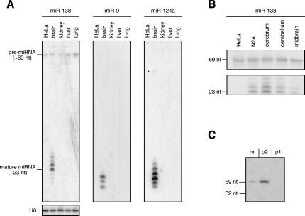
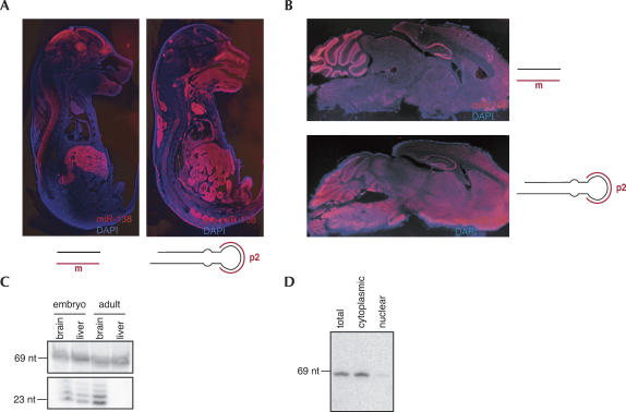
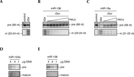
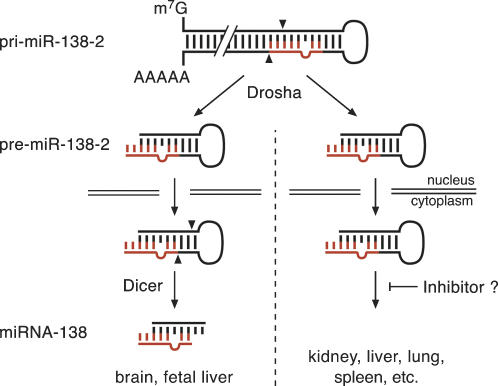
microRNAs (miRNAs) are endogenous, noncoding approximately 22-nucleotide RNA molecules that have recently emerged as fundamental, post-transcriptional regulators of cognate target gene expression. Many mammalian miRNAs are expressed in a tissue-specific manner, a phenomenon that has so far been attributed to transcriptional regulation. We here show by Northern blots and in situ hybridization experiments that the expression of mammalian miRNAs can be regulated at the post-transcriptional level. In particular, miR-138 is spatially restricted to distinct cell types, while its precursor, pre-miR-138-2, is ubiquitously expressed throughout all tissues analyzed. Furthermore, pre-miR-138-2 is exported from the nucleus to the cytoplasm, suggesting that cleavage of this pre-miRNA by Dicer is restricted to certain tissues and cell types. Thus, differential processing of pre-miRNAs might be an alternative mechanism to control miRNA function.


Figures

FIGURE 1.
FIGURE 1.
(A) Northern blot analysis of different brain-specific miRNAs. Thirty micrograms of total RNA from HeLa cells and from different mouse tissues were blotted and probed with a 5′-radiolabeled, LNA-modified oligodeoxynucleotide complementary to the indicated miRNAs. Equal loading of total RNA on the gel was verified by hybridizing with a probe complementary to the U6 snRNA. Note that only the precursor of miR-138 displays a ubiquitous pattern. The asterisk highlights the faint band of pre-miR-124a seen in the lane of murine brain at ∼57 nt. (B) Northern blot analysis of total RNA isolated from HeLa cells, from the murine neuroblastoma cell line N2A, and from different neuronal tissues of murine brain showing the tissue-specific processing of the ubiquitous 69-nt pre-miRNA. The band at 69 nt corresponds to the precursor, which is present in all cells and tissues that were analyzed. The 23- to 24-nt bands represent the mature miR-138, which is specifically expressed in the cerebrum as well as in the cerebellum, midbrain, and N2A cells, albeit to a lower extent. (C) Northern blot analysis of pre-miR-138-2 and pre-miR-138-1. One hundred micrograms of total RNA from murine brain were blotted and probed with 5′-radiolabeled, LNA-modified oligonucleotide probes complementary to the mature sequence of miR-138 (m), to the terminal loop of pre-miR-138-2 (p2), and to the terminal loop of pre-miR-138-1 (p1).
FIGURE 2.
FIGURE 2.
(A) In situ hybridization experiment on cryo-sections of E17 mouse embryos using LNA-modified probes that recognize mature miR-138 (left panel) or pre-miR-138-2 (right panel). The probe against mature miR-138 shows a strong expression in neuronal tissues (brain, CNS) and also in fetal liver, while the probe hybridizing to pre-miR-138-2 gives a strong signal in almost all tissues of the embryo. The seemingly low level of pre-miR-138-2 in brain can be explained by a low cellular density of the brain region as well as to a short exposure time used to prevent overexposure of tissues with higher cellular density. Magnifications of the fetal brain region can be seen in the Supplemental Material, where exposure time has been optimized to visualize expression of both mature miR-138 and pre-miR-138-2. (B) In situ hybridization experiments on cryo-sections of adult mouse brain using LNA-modified oligonucleotide probes that recognize mature miR-138 (upper panel) or pre-miR-138-2 (lower panel). miR-138 is primarily located to specific regions of the neocortex, most neurons in the hippocampus, and granule and purkinje cells of the cerebellum, while pre-miR-138-2 is essentially uniformly distributed. Magnifications of the cerebellum can be seen in the Supplemental Material. (C) Northern blot analysis of miR-138 expression in fetal and adult brain and liver. One hundred micrograms of total RNA prepared from the indicated tissues were blotted and probed for miR-138. In the mouse embryo, miR-138 is expressed in both brain and liver, which is in agreement with the findings in the in situ hybridization experiments. In the adult, miR-138 is restricted to brain. (D) Northern blot of total HeLa RNA and cytoplasmic and nuclear RNA fractions from HeLa cells probed against pre-miR-138-2. The band at 69 nt corresponds to pre-miR-138-2 that is mainly found in the cytoplasmic fraction, indicating nuclear export of the precursor.
FIGURE 3.
FIGURE 3.
(A) Processing reaction of pre-miR-138-2 into mature miR-138 by recombinant Dicer (rDcr) resolved on a 15% denaturing PAGE. (B) As in A. The addition of increasing amounts of HeLa cytoplasmic extract effectively abolishes the processing of pre-miR-138-2. The band corresponding to the respective pre-miRNA is indicated by “pre,” whereas the band representing the mature miRNA is designated by “m.” (C) Processing of pre-miR-19a into mature miR-19a is not abolished by addition of increasing amounts of HeLa cytoplasmic extract. The efficiency of processing is reduced by the addition of HeLa cytoplasmic extracts; however, the level of processing does not decrease with higher amounts of HeLa cytoplasmic extracts. (D) HeLa cells were transfected with the indicated amounts of a plasmid encoding the sequence of pre-miR-124a. The conversion of pre-miR-124a into mature miR-124a was monitored by Northern blotting. (E) HeLa cells were transfected with the indicated amounts of a plasmid encoding the sequence of pre-miR-138-2. In contrast to pre-miR-124a, HeLa cells failed to process pre-miR-138-2 into mature miR-138. Note that pre-miR-138-2 is more abundant than pre-miR-124a.
FIGURE 4.
FIGURE 4.
Model depicting how differential processing of an otherwise ubiquitously expressed pre-miRNA mediates tissue- and/or developmental stage-specific expression of the mature miRNA. In both tissues, expressing or not expressing the mature miR-138, its gene is transcribed into pri-miR-138-2, which is cleaved in the nucleus by Drosha into pre-miR-138-2 and then exported to the cytoplasm. Here, the presence or the absence of an inhibitory factor determines whether pre-miR-138-2 becomes processed or not, thus leading to differential expression of mature miR-138.

References

    1. Ambros V., Lee R.C., Lavanway A., Williams P.T., Jewell D. MicroRNAs and other tiny endogenous RNAs in C. elegans . Curr. Biol. 2003;13:807–818. - PubMed
    1. Babak T., Zhang W., Morris Q., Blencowe B.J., Hughes T.R. Probing microRNAs with microarrays: Tissue specificity and functional inference. RNA. 2004;10:1813–1819. - PMC - PubMed
    1. Barad O., Meiri E., Avniel A., Aharonov R., Barzilai A., Bentwich I., Einav U., Gilad S., Hurban P., Karov Y., et al. MicroRNA expression detected by oligonucleotide microarrays: System establishment and expression profiling in human tissues. Genome Res. 2004;14:2486–2494. - PMC - PubMed
    1. Bartel D.P. MicroRNAs: Genomics, biogenesis, mechanism, and function. Cell. 2004;116:281–297. - PubMed
    1. Bentwich I., Avniel A., Karov Y., Aharonov R., Gilad S., Barad O., Barzilai A., Einat P., Einav U., Meiri E., et al. Identification of hundreds of conserved and nonconserved human microRNAs. Nat. Genet. 2005;37:766–770. - PubMed

Publication types

LinkOut - more resources