. 2006:2:2006.0019.
doi: 10.1038/msb4100063. Epub 2006 May 2.

Residues crucial for maintaining short paths in network communication mediate signaling in proteins

Affiliations

Residues crucial for maintaining short paths in network communication mediate signaling in proteins

Antonio del Sol et al. Mol Syst Biol. 2006.

Abstract

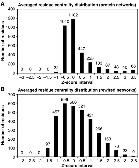
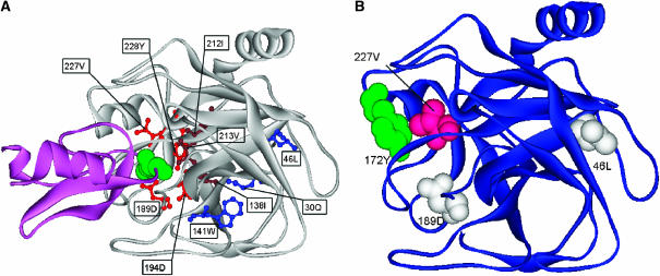
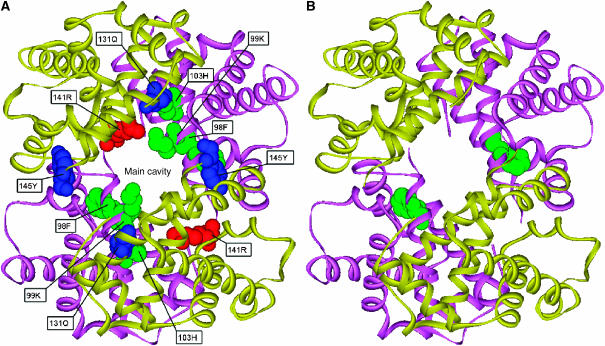
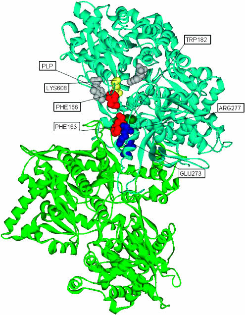
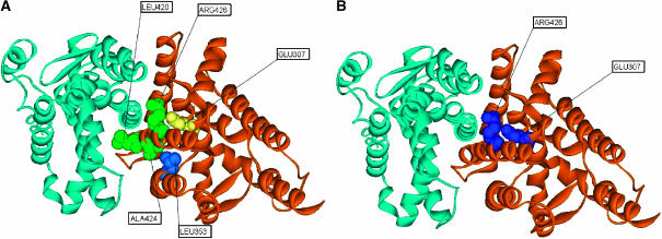
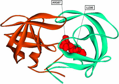
Here, we represent protein structures as residue interacting networks, which are assumed to involve a permanent flow of information between amino acids. By removal of nodes from the protein network, we identify fold centrally conserved residues, which are crucial for sustaining the shortest pathways and thus play key roles in long-range interactions. Analysis of seven protein families (myoglobins, G-protein-coupled receptors, the trypsin class of serine proteases, hemoglobins, oligosaccharide phosphorylases, nuclear receptor ligand-binding domains and retroviral proteases) confirms that experimentally many of these residues are important for allosteric communication. The agreement between the centrally conserved residues, which are key in preserving short path lengths, and residues experimentally suggested to mediate signaling further illustrates that topology plays an important role in network communication. Protein folds have evolved under constraints imposed by function. To maintain function, protein structures need to be robust to mutational events. On the other hand, robustness is accompanied by an extreme sensitivity at some crucial sites. Thus, here we propose that centrally conserved residues, whose removal increases the characteristic path length in protein networks, may relate to the system fragility.


Figures

Figure 1
Figure 1
(A) Averaged distribution of residue centrality of the seven representative protein structures. The average number of residues for each residue centrality z-score interval is indicated at the top of each bar. The average values of the network characteristic path length and clustering coefficient are L=4.21 and C=0.52, respectively. (B) Averaged distribution of residue centrality of the randomly rewired networks of the seven family representative protein structures. The average values of the network characteristic path length and clustering coefficient are L=2.62 and C=0.04, respectively.
Figure 2
Figure 2
Schematic representation of the analysis for determining the conserved central positions based on an example of a protein family comprising four proteins. The position shown in red in the family structural alignment is central in the network representation of each family member. In the family member structures, this same position is represented in blue.
Figure 3
Figure 3
Mapping of CICD residues onto the structure of the sperm whale myoglobin. The heme group is represented in brown and the atoms located at the xenon cavities are shown in green. Residues binding the heme group are shown in pink and blue, those lining the xenon cavities are colored pink and red and the redox-active residues are represented in yellow.
Figure 4
Figure 4
(A) CICD residues located in the three functional regions of the bovine rhodopsin structure. The cyclohexenyl ring of retinal is depicted in brown. Residues in blue are clustered at the bottom of the ligand-binding pocket, whereas those shown in red and green are located in the linking core and the G-protein-coupling region, respectively. (B) CID residues forming part of the network of coupling between positions in the GPCR family, as identified by Ranganathan and co-workers. CICD residues shown in red are part of the network of statistically coupled residues, whereas those represented in blue are neighbors of the residues colored in green belonging to this network.
Figure 5
Figure 5
(A) Structural mapping of CICD residues in the bovine beta-trypsin complex (gray) with pancreatic trypsin inhibitor (magenta). Residues belonging to the trypsin S1 pocket (red) are in contact with Lys15 of the pancreatic trypsin inhibitor (green). CICD residues (brown) located further from the binding site are likely to be important for the binding specificity, whereas those shown in blue reside in the core of the protein. (B) Correspondence between CICD residues and statistically coupled positions for trypsin, as detected by Ranganathan and co-workers. CICD residues (white) belong to the network of statistically coupled residues, whereas Val227 (pink) interacts with the statistically coupled residue Y172 (green).
Figure 6
Figure 6
(A) Representation of CICD residues in the structure of human hemoglobin. The two α and two β subunits are colored in magenta and yellow, respectively. CICD residues belonging to α subunits are located at the α1β12β2) interfaces (inside the hemoglobin central cavity, green) and at the interfaces α1β22β1) (red), whereas those from β subunits are part of the α1β12β2) and α1β22β1) interfaces (blue). (B) CICD residues forming part of the network of statistically coupled residues, as identified by Ranganathan and collaborators. The two α and two β subunits are colored in magenta and yellow, respectively. CICD residue Phe98 belonging to both α subunits is shown in green, and forms part of the network of coupled residues.
Figure 7
Figure 7
Mapping of CICD residues onto the homodimer structure of the rabbit muscle glycogen phosphorylase. The two subunits of the homodimer are represented in blue (chain A) and green (chain B). The PLP cofactor is represented in gray. Predicted residues binding the cofactor are colored in yellow. Residues located in one of the tower helices are shown in blue, those belonging to the beta turn are colored in red and the more hidden residue Trp182 is represented in dark green.
Figure 8
Figure 8
(A) CICD residues located in the structure of the homodimer human retinoic acid receptor RXR-alpha. The two subunits are represented in orange and blue, respectively. Residues forming part of the dimerization interface are depicted in green. Residue Glu307, important for the dimerization of LXR receptor, is colored in yellow. Residue Leu353, located between the ligand-binding site and the dimerization interface, is represented in blue. (B) The CICD amino acids corresponding to statistically coupled residues predicted by Ranganathan and co-workers are colored in blue.
Figure 9
Figure 9
Representation of CICD residues in the structure of HIV-1 protease complex (red color). Monomers are colored in blue and orange, respectively.

References

    1. Achacoso TB, Yamamoto WS (1992) AY's Neuroanatomy of C. elegans for Computation. Boca Raton, FL: CRC Press
    1. Aharoni A, Gaidukov L, Khersonsky O, Goulds MMcQ, Roodveldt C, Tawfik DS (2005) The evolvability of promiscuous protein functions. Nat Genet 37: 73–76 - PubMed
    1. Amitai G, Shemesh A, Sitbon E, Shklar M, Netanely D, Venger I, Pietrokovski S (2004) Network analysis of protein structures identifies functional residues. J Mol Biol 344: 1135–1146 - PubMed
    1. Andres A, Kosoy A, Garriga P, Manyosa J (2001) Mutations at position 125 in transmembrane helix III of rhodopsin affect the structure and signalling of the receptor. Eur J Biochem 268: 5696–5704 - PubMed
    1. Ballesteros JA, Shi L, Javitch JA (2001) Structural mimicry in G protein-coupled receptors: implications of the high-resolution structure of rhodopsin for structure–function analysis of rhodopsin-like receptors. Mol Pharmacol 60: 1–19 - PubMed

Publication types