. 2006 Jun 1;7(1):85.
doi: 10.1186/1465-9921-7-85.

Susceptibility to ozone-induced airway inflammation is associated with decreased levels of surfactant protein D

Affiliations

Susceptibility to ozone-induced airway inflammation is associated with decreased levels of surfactant protein D

S Kierstein et al. Respir Res. .

Abstract

Background: Ozone (O3), a common air pollutant, induces exacerbation of asthma and chronic obstructive pulmonary disease. Pulmonary surfactant protein (SP)-D modulates immune and inflammatory responses in the lung. We have shown previously that SP-D plays a protective role in a mouse model of allergic airway inflammation. Here we studied the role and regulation of SP-D in O3-induced inflammatory changes in the lung.

Methods: To evaluate the effects of O3 exposure in mouse strains with genetically different expression levels of SP-D we exposed Balb/c, C57BL/6 and SP-D knockout mice to O3 or air. BAL cellular and cytokine content and SP-D levels were evaluated and compared between the different strains. The kinetics of SP-D production and inflammatory parameters were studied at 0, 2, 6, 12, 24, 48, and 72 hrs after O3 exposure. The effect of IL-6, an O3-inducible cytokine, on the expression of SP-D was investigated in vitro using a primary alveolar type II cell culture.

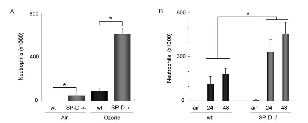
Results: Ozone-exposed Balb/c mice demonstrated significantly enhanced acute inflammatory changes including recruitment of inflammatory cells and release of KC and IL-12p70 when compared with age- and sex-matched C57BL/6 mice. On the other hand, C57BL/6 mice had significantly higher levels of SP-D and released more IL-10 and IL-6. Increase in SP-D production coincided with the resolution of inflammatory changes. Mice deficient in SP-D had significantly higher numbers of inflammatory cells when compared to controls supporting the notion that SP-D has an anti-inflammatory function in our model of O3 exposure. IL-6, which was highly up-regulated in O3 exposed mice, was capable of inducing the expression of SP-D in vitro in a dose dependent manner.

Conclusion: Our data suggest that IL-6 contributes to the up-regulation of SP-D after acute O3 exposure and elevation of SP-D in the lung is associated with the resolution of inflammation. Absence or low levels of SP-D predispose to enhanced inflammatory changes following acute oxidative stress.


Figures

Figure 1
Figure 1
A decrease in BAL SP-D levels was associated with significantly increased inflammation in Balb/c mice 6 hrs after O3 exposure. Groups of Balb/c (grey bars) and C57BL/6 (black bars) mice were exposed to O3 or room air for 2 hrs and BAL was performed 6 hrs later. (A) Ozone exposed Balb/c mice had significantly reduced SP-D levels as compared to air exposed Balb/c or O3 exposed C57BL/6 mice. SP-D was detected by Western blot analysis of the cell-free supernatant of the BAL (top panel) and was performed using our in-house rabbit polyclonal anti-SP-D antibody. Two representative samples out of a total of six are shown in each group. SP-D expression was quantified by densitometric analysis. Results are expressed as % of naïve control levels. * p = 0.0249 vs. room air; p = 0.0027 vs. C57BL/6. (B) Balb/c mice had significantly higher numbers of inflammatory cells in their BAL as compared to C57BL/6 mice. Cells were counted using a Coulter counter and results are expressed as cell number/ml (*p = 0.0316). (C): The BAL total protein content was significantly higher in Balb/c mice compared to C57BL/6 mice. Total protein was measured by Bradford assay in the cell-free supernatant *p = 0.028. Absolute protein contents were 919,6 (± 51,4) and 3262,3 (± 281.0) in air and O3 exposed Balb/c mice, respectively, and 772 (± 26.4) and 2110 (± 36.4) in air and O3 exposed C57BL/6 mice, respectively.(D-G): Cytokine expression was studied by Endogen®SearchLight™ and Luminex®100™ technologies. O3 induced the release of IL-12p70 (34 pg/ml ± 4 in Balb/c, 18.7 pg/ml ± 1 in C57BL/6), IL-6 (2,393 pg/ml ± 119 in Balb/c, 412 pg/ml ± 68.7 in C57BL/6), IL-10 (110 pg/ml ± 16 in Balb/c, 14.2 pg/ml ± 1.6 in C57BL/6) and KC (1,896 pg/ml ± 224 in Balb/c, 136.8 pg/ml ± 27.7 in C57BL/6). Cytokine and chemokine levels are expressed as % increase from control levels. The O3- induced pro-inflammatory cytokine IL-12p70 and KC levels were significantly higher (*p = 0.0134 and *p = 0.0001, respectively) whereas the immunosuppressive IL-10 and the immunoregulatory IL-6 levels were significantly lower (*p < 0.0001 and *p < 0.0001, respectively) in Balb/c mice than in C57BL/6 mice. (A-G): Mean ± SEM of n = 6 in each groups.
Figure 2
Figure 2
Kinetics of O3-induced SP-D and inflammatory changes in C57BL/6 mice. Groups of C57BL/6 mice were exposed to 3 ppm O3 or room air and studied 0, 2, 6, 12, 24, 48, and 72 hrs later. (A): SP-D and SP-A recovered from the BAL were quantified by an in-house ELISA. Results are expressed as % change from naïve controls. SP-D levels gradually increased until the 72 hrs time point (p = 0.0022, ANOVA and post test for linear trend) whereas SP-A levels did not change significantly. (B): The number of neutrophilic granulocytes was assessed by counting total number of BAL cells in the BAL fluid and performing differential cell counting on Kwick™Diff cytospin preparations. Results are expressed as absolute cell number/ml of BAL fluid. Neutrophilic inflammation peaked around 12 hrs post exposure (p < 0.0001) and largely resolved by 72 hrs after O3 exposure. (C-D) IL-6 and IL-10 levels were assessed as part of a Luminex®100™ assay and showed significant increases 2–6 hrs after O3 exposure (p < 0.0001) and 6–24 hrs after O3 exposure (p = 0.0007), respectively. (A-D): Mean ± SEM of n = 6 in each groups.
Figure 3
Figure 3
SP-D deficient mice have increased airway inflammation following O3 exposure. (A) SP-D -/- mice and age-matched C57BL/6 controls were exposed to 3 ppm O3 for 2 hrs or (B) to 0.5 ppm for 24 hrs. Influx of neutrophilic granulocytes was assessed on cytospin preparations stained with Kwick™Diff. In both models cellular inflammation in SP-D -/- mice was significantly higher compared to wt mice (A) Student t-test *p = 0.0106 (B) ANOVA and Barlett's test *p = 0.0082.
Figure 4
Figure 4
In vitro production of SP-D by alveolar type II cells after IL-6 stimulation. (A) Primary rat alveolar type II cells were cultured in the presence or absence of DCI and IL-6 for up to 4 days and intra-cellular SP-D and SP-A levels were assayed by Western blot analysis. Day 0 SP-D and SP-A signals were obtained from freshly isolated alveolar type II cells. To maintain the SP-D producing phenotype, cells have to be cultured in the presence of DCI (10 nM dexamethason, 100 nM IBMX, 10 nM cAMP) that in turn stimulates SP-D production. IL-6 selectively stimulated the production of SP-D but did not induce up-regulation of SP-A. (B) Increase of SP-D after IL-6 stimulation at day 4 was slightly dose dependent. All levels were compared to non-stimulated alveolar type II cells cultured in DCI containing medium (DCI control).

References

    1. Desqueyroux H, Pujet JC, Prosper M, Le Moullec Y, Momas I. Effects of air pollution on adults with chronic obstructive pulmonary disease. Arch Environ Health. 2002;57(6):554–560. - PubMed
    1. Peel JL, Tolbert PE, Klein M, Metzger KB, Flanders WD, Todd K, Mulholland JA, Ryan PB, Frumkin H. Ambient air pollution and respiratory emergency department visits. Epidemiology. 2005;16(2):164–174. doi: 10.1097/01.ede.0000152905.42113.db. - DOI - PubMed
    1. Stenfors N, Pourazar J, Blomberg A, Krishna MT, Mudway I, Helleday R, Kelly FJ, Frew AJ, Sandstrom T. Effect of ozone on bronchial mucosal inflammation in asthmatic and healthy subjects. Respir Med. 2002;96(5):352–358. doi: 10.1053/rmed.2001.1265. - DOI - PubMed
    1. Savov JD, Whitehead GS, Wang J, Liao G, Usuka J, Peltz G, Foster WM, Schwartz DA. Ozone-induced acute pulmonary injury in inbred mouse strains. Am J Respir Cell Mol Biol. 2004;31(1):69–77. doi: 10.1165/rcmb.2003-0001OC. - DOI - PubMed
    1. McKinney WJ, Jaskot RH, Richards JH, Costa DL, Dreher KL. Cytokine mediation of ozone-induced pulmonary adaptation. Am J Respir Cell Mol Biol. 1998;18(5):696–705. - PubMed

MeSH terms