. 2006 Jun 1:6:90.
doi: 10.1186/1471-2334-6-90.

Vaginal microbicides: detecting toxicities in vivo that paradoxically increase pathogen transmission

Affiliations

Vaginal microbicides: detecting toxicities in vivo that paradoxically increase pathogen transmission

Richard A Cone et al. BMC Infect Dis. .

Abstract

Background: Microbicides must protect against STD pathogens without causing unacceptable toxic effects. Microbicides based on nonoxynol-9 (N9) and other detergents disrupt sperm, HSV and HIV membranes, and these agents are effective contraceptives. But paradoxically N9 fails to protect women against HIV and other STD pathogens, most likely because it causes toxic effects that increase susceptibility. The mouse HSV-2 vaginal transmission model reported here: (a) Directly tests for toxic effects that increase susceptibility to HSV-2, (b) Determines in vivo whether a microbicide can protect against HSV-2 transmission without causing toxicities that increase susceptibility, and (c) Identifies those toxic effects that best correlate with the increased HSV susceptibility.

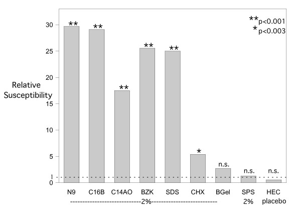
Methods: Susceptibility was evaluated in progestin-treated mice by delivering a low-dose viral inoculum (0.1 ID50) at various times after delivering the candidate microbicide to detect whether the candidate increased the fraction of mice infected. Ten agents were tested - five detergents: nonionic (N9), cationic (benzalkonium chloride, BZK), anionic (sodium dodecylsulfate, SDS), the pair of detergents in C31G (C14AO and C16B); one surface active agent (chlorhexidine); two non-detergents (BufferGel, and sulfonated polystyrene, SPS); and HEC placebo gel (hydroxyethylcellulose). Toxic effects were evaluated by histology, uptake of a 'dead cell' dye, colposcopy, enumeration of vaginal macrophages, and measurement of inflammatory cytokines.

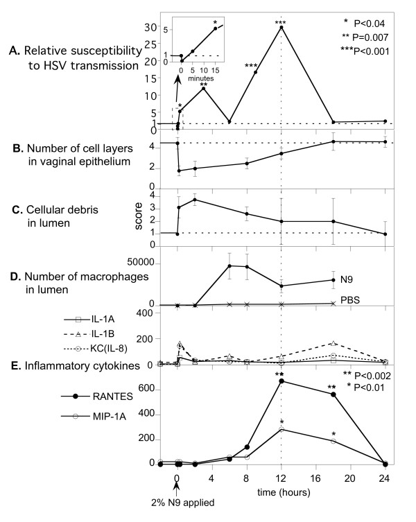
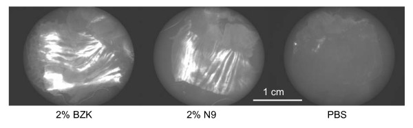
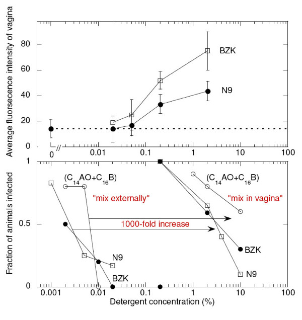
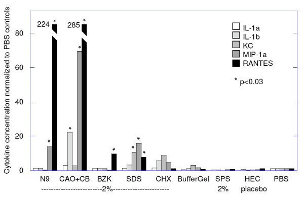
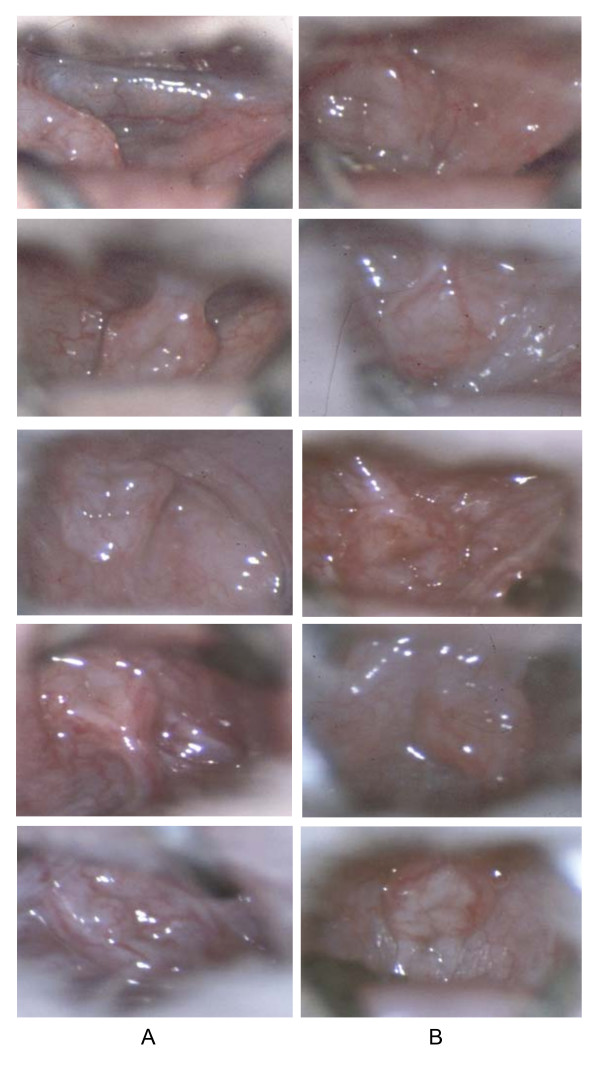
Results: A single dose of N9 protected against HSV-2 for a few minutes but then rapidly increased susceptibility, which reached maximum at 12 hours. When applied at the minimal concentration needed for brief partial protection, all five detergents caused a subsequent increase in susceptibility at 12 hours of approximately 20-30-fold. Surprisingly, colposcopy failed to detect visible signs of the N9 toxic effect that increased susceptibility at 12 hours. Toxic effects that occurred contemporaneously with increased susceptibility were rapid exfoliation and re-growth of epithelial cell layers, entry of macrophages into the vaginal lumen, and release of one or more inflammatory cytokines (Il-1beta, KC, MIP 1alpha, RANTES). The non-detergent microbicides and HEC placebo caused no significant increase in susceptibility or toxic effects.

Conclusion: This mouse HSV-2 model provides a sensitive method to detect microbicide-induced toxicities that increase susceptibility to infection. In this model, there was no concentration at which detergents provided protection without significantly increasing susceptibility.


Figures

Figure 1
Figure 1
Vaginal HSV-2 susceptibility and toxic effects vs time after a single dose of 2% N9. A. Relative susceptibility is the effective Infectious Doses the inoculum delivers in N9-exposed mice normalized to the Infectious Doses it delivers in control (PBS) mice. The single dose of N9 produced three successive phases in susceptibility, a brief initial protective phase (reduced susceptibility) followed by two distinct phases of markedly increased susceptibility. P values were determined by Fisher's exact two-sided test comparing the numbers of animals infected and uninfected in the test animals vs. the corresponding numbers in control animals treated only with PBS. B-E. Error bars indicate standard deviations around the mean. Concentrations of inflammatory cytokines are indicated by geometric means normalized with respect to those for PBS controls. See legend for Fig. 6 for geometric means of PBS controls. B, C. Data obtained from histology sections from four locations distributed along the length of the vagina. The score in C is a semi-quantitative evaluation of the amount of visible cellular debris in the vaginal lumen. For each point in B and C at 10 min, 2, and 8 hours, P values were <0.001 with respect to PBS control values. (n = 8) D. P values for all times points from 6 to 18 hours were <0.001 with respect to PBS control values. (n = 20) D, E. Data obtained from vaginal lavage samples.
Figure 2
Figure 2
Vaginal HSV-2 susceptibility 12 hours after a single application of candidate microbicides. P values (from Table 1), determined by Fisher's exact two-sided test as described in the caption for Figure 1. n.s. = not significant.
Figure 3
Figure 3
A single previous exposure to N9 can abolish the brief protective effect of N9. The low-dose inoculum (0.05 ID50) infected only a small fraction of the control group treated twice with PBS (first column), and an even smaller fraction was infected in the control group treated with 2% N9 just before delivering the inoculum (middle column). But a significantly larger fraction became infected in the test group (right-hand column), revealing that the N9 dose delivered 12-hour earlier abolished the protective effect of the N9 dose delivered just before the inoculum: P = 0.013 by Fisher's exact two-sided test.
Figure 4
Figure 4
Surface distribution of acute toxicity to vaginal epithelium caused by a 10-minute exposure to detergents. Fluorescent intensity of the 'dead cell' dye YOYO-1 is proportional to the density of cell nuclei in the epithelium whose membranes were disrupted by the detergents. The brightly stained swaths of epithelium are regions in which virtually every cell nuclei became stained with YOYO-1. The darker regions became visible as the vagina was gently stretched and flattened to unfold the rugae.
Figure 5
Figure 5
Acute toxicity, and fraction of mice infected, vs detergent concentration. The upper panel shows the average fluorescent intensity of the YOYO-1 stained vagina as a function of the concentration of detergent to which each vagina was exposed for 10 minutes. The lower panel shows the fraction of mice infected using two different test methods: 'mix externally' fully exposes HSV in a high-dose inoculum (10 ID50) to the microbicide prior to inoculation, and 'mix in vagina' mimics human use of a microbicide, the microbicide is delivered to the vagina before delivering the high-dose (10 ID50) inoculum. The two detergents in C31G, (C14AO+C16B), were combined at equal w/v concentrations and plotted at the total detergent concentrations shown.
Figure 6
Figure 6
Inflammatory cytokines 12 hours after a single exposure to candidate microbicides. Geometric means for each agent are shown normalized by the results in PBS controls as follows, in ng/ml of lavage fluid: IL-1α, 16; IL-1β, 50; KC, 42; MIP-1α, 20; RANTES, 3. Thus N9 increased the concentration of RANTES 224-fold, from 3 to 672 ng/ml in lavage fluid. (The lavage procedure is estimated to have diluted vaginal secretions by 10–50-fold.) (CAO+CB) = 1%CAO+1%CB = 2% detergent.
Figure 7
Figure 7
Colposcopic images of mouse vaginas 12 hours after a single exposure to 2% N9, or PBS. The colposcope (Zeiss OPMI 1-SH) was set at maximum magnification (31×) and focused on the vaginal area surrounding the cervix. Column A, 2% N9, column B, PBS controls.

References

    1. Sodora DL, Gettie A, Miller CJ, Marx PA. Vaginal transmission of SIV: assessing infectivity and hormonal influences in macaques inoculated with cell-free and cell-associated viral stocks. AIDS Res Hum Retroviruses. 1998;14:S119–123. - PubMed
    1. Chakraborty H, Sen PK, Helms RW, Vernazza PL, Fiscus SA, Eron JJ, Patterson BK, Coombs RW, Krieger JN, Cohen MS. Viral burden in genital secretions determines male-to-female sexual transmission of HIV-1: a probabilistic empiric model. Aids. 2001;15:621–627. doi: 10.1097/00002030-200103300-00012. - DOI - PubMed
    1. Wawer MJ, Gray RH, Sewankambo NK, Serwadda D, Li X, Laeyendecker O, Kiwanuka N, Kigozi G, Kiddugavu M, Lutalo T, et al. Rates of HIV-1 transmission per coital act, by stage of HIV-1 infection, in Rakai, Uganda. J Infect Dis. 2005;191:1403–1409. doi: 10.1086/429411. - DOI - PubMed
    1. Catalone BJ, Miller SR, Ferguson ML, Malamud D, Kish-Catalone T, Thakkar NJ, Krebs FC, Howett MK, Wigdahl B. Toxicity, inflammation, and anti-human inmmunodeficiency virus type I activity following exposure to chemical moieties of C31G. Biomedicine & Pharmacotherapy. 2005;59:430–437. doi: 10.1016/j.biopha.2005.07.008. - DOI - PubMed
    1. Catalone BJ, Kish-Catalone TM, Budgeon LR, Neely EB, Ferguson M, Krebs FC, Howett MK, Labib M, Rando R, Wigdahl B. Mouse model of cervicovaginal toxicity and inflammation for preclinical evaluation of topical vaginal microbicides. Antimicrob Agents Chemother. 2004;48:1837–1847. doi: 10.1128/AAC.48.5.1837-1847.2004. - DOI - PMC - PubMed

Publication types

MeSH terms