Review
. 2006 Aug;98(2):279-88.
doi: 10.1093/aob/mcl107. Epub 2006 Jun 1.

Could heat shock transcription factors function as hydrogen peroxide sensors in plants?

Affiliations
Review

Could heat shock transcription factors function as hydrogen peroxide sensors in plants?

Gad Miller et al. Ann Bot. 2006 Aug.

Abstract

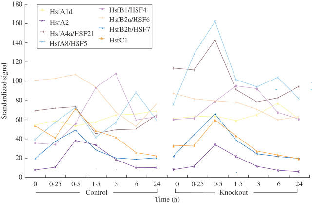
Background: Heat shock transcription factors (Hsfs) are modular transcription factors encoded by a large gene family in plants. They bind to the consensus sequence 'nGAAnnTCCn' found in the promoters of many defence genes, and are thought to function as a highly redundant and flexible gene network that controls the response of plants to different environmental stress conditions, including biotic and abiotic stresses. Hsf proteins encoded by different genes exhibit a high degree of complexity in their interactions. They can potentially bind and activate their own promoters, as well as the promoters of other members of their gene family, and they can form homo- or heterotrimers resulting in altered nuclear localization, as well as enhanced or suppressed transcription.

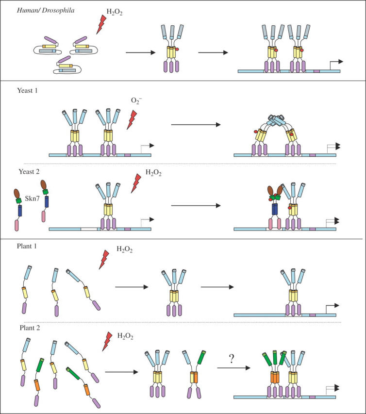
Scope: In this review, we summarize recent studies on Hsf function in Arabidopsis and tomato and present evidence obtained from microarray expression studies in Arabidopsis that the Hsf gene network is highly flexible and specialized, with specific members and/or member combinations controlling the response of plants to particular stress conditions. In addition, we describe recent studies that support the hypothesis that certain Hsfs function as molecular sensors that directly sense reactive oxygen species (ROS) and control the expression of oxidative stress response genes during oxidative stress.


Figures

F<sc>ig</sc>. 1.
Fig. 1.
Arabidopsis Hsf protein structure. A model representing the structure of Arabidopsis HsfA1a (A-Class Hsf), HsfB1 (B-Class Hsf) and HsfC1 (C-Class Hsf) is shown. The different domains are indicated on the top and different structural features present in each domain are outlined below. The models presented in the figure were generated according to Nover et al. (2001).
F<sc>ig</sc>. 2.
Fig. 2.
Steady-state transcript level of Arabidopsis HSFs in different tissues. The basal steady-state transcript level of all Arabidopsis Hsfs was obtained from the Genevestigator microarray database using the ‘Meta-analyzer’ tool (Zimmermann et al., 2004). The values represent the signal intensity of each probe set as given by Genevestigator (https://www.genevestigator.ethz.ch/).
F<sc>ig</sc>. 3.
Fig. 3.
Arabidopsis Hsf transcript accumulation in response to different environmental stress conditions. The values are presented as fold change, relative to control treatment or t = 0 min of the experiment. All data, except for the light stress experiment, were obtained from the Genevestigator microarray database using the ‘digital-northern’ tool (Zimmermann et al., 2004). The relative induction levels of Hsfs during moderate light stress were obtained from a microarray experiment previously described by Davletova et al. (2005a). To demonstrate maximal fold increase for each Hsf transcript in each stress treatment, in each experiment that contained several time points, the time point in which a particular Hsf showed the highest expression level was chosen for representation. In general, the following time points were used: light stress (30 min), salt (12 h), drought (24 h), cold (24 h) and wounding (24 h). The following exceptions were made for particular Hsfs showing the highest level of induction at other time points: Light stress: HsfB1, 3 h; HsfA8, 6 h. Salt stress: HsfA1a, HsfA4a, HsfA6a, HsfB2a, 6 h; A1e, A8, A9, B1, B2b and B3, 24 h. Cold stress: A4a and B2a, 6 h; A1d and A5, 12 h. Wounding: B1 and 6a, 6 h.
F<sc>ig</sc>. 4.
Fig. 4.
A putative model for Hsf function in different organisms. The human and Drosophila Hsfs (upper panel) are inactive under non-stress conditions due to intramolecular interactions between HR-A/B and the HR-C domains. In response to hydrogen peroxide, the protein forms a disulfide bond between two cysteine residues inside and near the DNA-binding domain. The active Hsf is then phosphorylated and forms a transcriptionaly active homotrimer that is transported to the nucleus and activates ROS-responsive gene expression. Two different models are presented for oxidative stress-mediated activation of Hsf in yeast (middle panels). In the upper section, both Hsfs are bound to the HSEs of the target gene promoter as a homotrimer; superoxide anions directly interact with the protein inducing a conformational change that forms a cooperative interaction between them that increases their transcriptional activity. The second yeast model (lower section) shows the increase of Hsf activity by the co-activation of Skn7. Upon perception of H2O2 stress, Skn7, which is localized in the nucleus, is phosphorylated at the receiver domain by a histidine kinase sensor inducing the formation of an Skn7 homodimer. The active homodimer can bind to HSE adjacent to Hsf and increase its transcriptional activation. Two hypothetical models for plants are presented in the lower panels. A simplistic model is shown in the upper section in which Hsf (AtHsfA4a or AtHsfA2a) forms a homotrimer in response to interaction with ROS and is transported to the nucleus to activate oxidative stress gene expression. In the lower section, oxidative stress induces the homo-trimerization of a particular Hsf, which in response interacts with another Hsf to mediate its transport to the nucleus. The two active Hsfs can cooperatively induce gene expression. For example, AtHsfA8a could act as the co-activator. AtHsfA8a is expressed during oxidative stresses (Fig. 3), and in KO-Apx1 plants (see Fig. 5). It is localized to the cytoplasm and it could be dependent on other Hsfs for nuclear localization. Alternatively, the homotrimer of AtHsfA4a can be transported to the nucleus where it can cooperate with a class B Hsf (AtHsfB2b) or another class A Hsf.
F<sc>ig</sc>. 5.
Fig. 5.
Time course analysis of Arabidopsis Hsf accumulation in response to light stress in wild-type and knockout plants lacking the ROS defence enzyme cytosolic ascorbate peroxidase (KO-Apx1). The expression of the eight light stress-responsive Hsfs is presented, in the wild type (left) and KO-Apx1 (right) during time course exposure to moderate light stress. The steady-state transcripts level was obtained from microarray experiments previously described by Daveltova et al. (2005a).

References

    1. Ahn SG, Thiele DJ. 2003. Redox regulation of mammalian heat shock factor 1 is essential for Hsp gene activation and protection from stress. Genes and Development 17: 516–528. - PMC - PubMed
    1. Apel K, Hirt H. 2004. Reactive oxygen species: metabolism, oxidative stress, and signal transduction. Annual Review of Plant Biology 55: 373–399. - PubMed
    1. Baniwal SK, Bharti K, Chan KY, Fauth M, Ganguli A, Kotak S, et al.2004. Heat stress response in plants: a complex game with chaperones and more than twenty heat stress transcription factors. Journal of Biosciences 29: 471–487. - PubMed
    1. Batulan Z, Shinder GA, Minotti S, He BP, Doroudchi MM, Nalbantoglu J, Strong MJ, Durham HD. 2003. High threshold for induction of the stress response in motor neurons is associated with failure to activate Hsf1. Journal of Neuroscience 23: 5789–5798. - PMC - PubMed
    1. Bergmüller E, Porfirova E, Dörmann P. 2003. Characterization of an Arabidopsis mutant deficient in γ-tocopherol methyltransferase. Plant Molecular Biology 52: 1181–1190. - PubMed

Publication types

MeSH terms