. 2006 Jun;188(12):4264-70.
doi: 10.1128/JB.00004-06.

RcsF is an outer membrane lipoprotein involved in the RcsCDB phosphorelay signaling pathway in Escherichia coli

Affiliations

RcsF is an outer membrane lipoprotein involved in the RcsCDB phosphorelay signaling pathway in Escherichia coli

Marie-Pierre Castanié-Cornet et al. J Bacteriol. 2006 Jun.

Abstract

The RcsCDB signal transduction system is an atypical His-Asp phosphorelay conserved in gamma-proteobacteria. Besides the three proteins directly involved in the phosphorelay, two proteins modulate the activity of the system. One is RcsA, which can stimulate the activity of the response regulator RcsB independently of the phosphorelay to regulate a subset of RcsB targets. The other is RcsF, a putative outer membrane lipoprotein mediating the signaling to the sensor RcsC. How RcsF transduces the signal to RcsC is unknown. Although the molecular and physiological signals remain to be identified, the common feature among the reported Rcs-activating conditions is perturbation of the envelope. As an initial step to explore the RcsF-RcsC functional relationship, we demonstrate that RcsF is an outer membrane lipoprotein oriented towards the periplasm. We also report that a null mutation in surA, a gene required for correct folding of periplasmic proteins, activates the Rcs pathway through RcsF. In contrast, activation of this pathway by overproduction of the membrane chaperone-like protein DjlA does not require RcsF. Conversely, activation of the pathway by RcsF overproduction does not require DjlA either, indicating the existence of two independent signaling pathways toward RcsC.


Figures

FIG. 1.
FIG. 1.
Absence of SurA activates the Rcs pathway in an RcsC- and an RcsF-dependent manner. Strains carrying an rprA-lacZ fusion (A) and strains carrying a cps-lacZ fusion (B) were assayed for β-galactosidase activity as described in Materials and Methods. Bars represent the standard deviations of at least three independent measurements. (See Table 1 for complete genotypes of strains.) Relevant genotypes are indicated under each measurement.
FIG. 2.
FIG. 2.
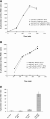
DjlA and RcsF act independently. Induction of cps-lacZ fusion expression by DjlA (A) or by RcsF (B) overproduction. Strains GEB495 (cps-lacZ), GEB496 (djlA::spc cps-lacZ), and GEB628 (rcsF::cat sacB cps-lacZ) were transformed with either pPSG958, prcsF, or pIM10 plasmid DNA. Cells were cultured overnight in LB in the presence of the appropriate antibiotic and then diluted to 1/1,000 into fresh LB and grown at 30°C. At an OD600 of approximately 0.1, 500 μM IPTG was added to some cultures, while others were left untreated. The cultures were sampled at regular intervals for assay of β-galactosidase activities. (C) The djlA::spc allele was introduced into GEB495 (cps-lacZ) and GEB498 (cps-lacZ surA::kan), and the resulting strains were assayed for β-galactosidase activity as described in Materials and Methods.
FIG. 3.
FIG. 3.
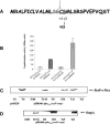
Signal sequence and localization of RcsF-c-Myc. (A) Signal sequence of RcsF. The predicted cleavage site, the acylated cysteine residue at position +1, and the serine residue at position +2 are indicated. (B) Complementation of rcsF mutant by RcsF-c-Myc. rprA-lacZ rcsF mutant and rprA-lacZ rcsF surA double mutant strains, transformed with either pAM238 (vector) or pHK646 (encoding RcsF-c-Myc), were assayed for β-galactosidase activity. (C and D) Cells from strain GEB658 (rprA-lacZ rcsF+) transformed with either pAM238 or pHK646 were fractionated as described in Materials and Methods, and the fractions from GEB658/pHK646 were analyzed for the presence of RcsF-c-Myc by Western blotting (C). Ttl, total fraction (whole cells); S100, soluble cytoplasmic and periplasmic content; Ttl M, total membrane fraction; IM, inner membrane fraction; OM, outer membrane fraction; TxS, Triton X-100-solubilized total membrane fraction; TxI, Triton X-100-insoluble total membrane fraction. (D) The same fractions were analyzed by Western blotting for the presence of OmpA.
FIG. 4.
FIG. 4.
Inhibition of processing of RcsF-c-Myc by globomycin. Strain 61F/pHK646 was grown in LB supplemented with spectinomycin. RcsF-c-Myc production was induced with IPTG in some cultures and left uninduced in others, and some cultures were treated with globomycin while others were not. Proteins from each culture were separated by SDS-PAGE. After transfer, the upper part of the membrane was immunoblotted with SurA antibodies as a loading control (upper panel), and the bottom part of the membrane was immunoblotted with c-Myc-specific monoclonal antibodies (lower panel; see Materials and Methods). The upper band detected in globomycin-treated cultures, indicated by an asterisk, corresponds to the unprocessed RcsF-c-Myc precursor.
FIG. 5.
FIG. 5.
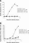
RcsF-PhoA is periplasmic. MP110 (cps-lacZ) strains transformed either with prcsF-phoA or prcsF-phoAΔ20 were grown overnight in LB in the presence of spectinomycin and then diluted 1,000-fold in fresh LB and grown to an OD600 of ∼0.1 at 37°C. At time zero, 0.2% arabinose was added to some cultures but not to others, and the cultures were sampled at regular intervals to assay either for alkaline phosphatase activity (A) or for β-galactosidase activity (B).

References

    1. Behrens, S. 2002. Periplasmic chaperones-new structural and functional insights. Structure (Cambridge) 10:1469-1471. - PubMed
    1. Bernard, S., D. J. Clarke, M. X. Chen, I. B. Holland, and A. Jacq. 1998. Increased sensitivity of E. coli to novobiocin, EDTA and the anticalmodulin drug W7 following overproduction of DjlA requires a functional transmembrane domain. Mol. Gen. Genet. 259:645-655. - PubMed
    1. Clarke, D. J., I. B. Holland, and A. Jacq. 1997. Point mutations in the transmembrane domain of DjlA, a membrane-linked DnaJ-like protein, abolish its function in promoting colanic acid production via the Rcs signal transduction pathway. Mol. Microbiol. 25:933-944. - PubMed
    1. Clarke, D. J., A. Jacq, and I. B. Holland. 1996. A novel DnaJ-like protein in Escherichia coli inserts into the cytoplasmic membrane with a type III topology. Mol. Microbiol. 20:1273-1286. - PubMed
    1. Clarke, D. J., S. A. Joyce, C. M. Toutain, A. Jacq, and I. B. Holland. 2002. Genetic analysis of the RcsC sensor kinase from Escherichia coli K-12. J. Bacteriol. 184:1204-1208. - PMC - PubMed

Publication types

MeSH terms

LinkOut - more resources