. 2006 Jun;188(12):4331-9.
doi: 10.1128/JB.00005-06.

Borrelia burgdorferi EbfC, a novel, chromosomally encoded protein, binds specific DNA sequences adjacent to erp loci on the spirochete's resident cp32 prophages

Affiliations

Borrelia burgdorferi EbfC, a novel, chromosomally encoded protein, binds specific DNA sequences adjacent to erp loci on the spirochete's resident cp32 prophages

Kelly Babb et al. J Bacteriol. 2006 Jun.

Abstract

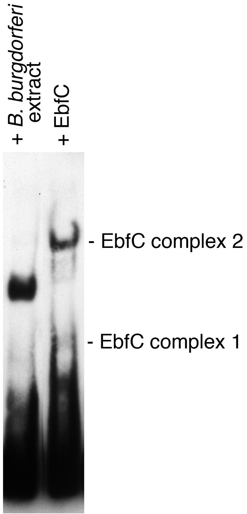
All examined isolates of the Lyme disease spirochete, Borrelia burgdorferi, naturally maintain numerous variants of a prophage family as circular cp32 episomes. Each cp32 carries a locus encoding one or two different Erp outer membrane, surface-exposed lipoproteins. Many of the Erp proteins bind a host complement regulator, factor H, which is hypothesized to protect the spirochete from complement-mediated killing. We now describe the isolation and characterization of a novel, chromosomally encoded protein, EbfC, that binds specific DNA sequences located immediately 5' of all erp loci. This is one of the first site-specific DNA-binding proteins to be identified in any spirochete. The location of the ebfC gene on the B. burgdorferi chromosome suggests that the cp32 prophages have evolved to use this bacterial host protein for their own benefit and that EbfC probably plays additional roles in the bacterium. A wide range of other bacteria encode homologs of EbfC, none of which have been well characterized, so demonstration that B. burgdorferi EbfC is a site-specific DNA-binding protein has broad implications across the eubacterial kingdom.


Figures

FIG. 1.
FIG. 1.
Alignment of noncoding DNA sequences located immediately 5′ of the determined erp loci of B. burgdorferi strains B31, N40, and 297 (the erp loci of strain Sh-2-82 are identical to those of strain 297 plus the erpNO locus of strain B31). Identical nucleotides found in the majority of loci are boxed and shaded. Initiation methionine codons of the first gene in each locus are to the far right. The maximum size of erp operator 2, as determined previously using competitive EMSA and transcriptional fusions to reporter genes (7), is indicated. The two EbfC-binding sites (TGT[A/T]ACA) determined by the present work are indicated as Site I and Site II above the alignment.
FIG. 2.
FIG. 2.
B. burgdorferi cytoplasmic proteins purified by affinity chromatography using erpG 5′ noncoding DNA as bait. Proteins in elutions 1, 2, and 3 were eluted with NaCl at concentrations of 500, 750, and 1,000 mM, respectively. Proteins were separated by SDS-PAGE and visualized with SYPRO-Ruby. Numbers on the right indicate positions of molecular mass standards.
FIG. 3.
FIG. 3.
Alignment of the predicted amino acid sequences of B. burgdorferi EbfC and a homologous protein encoded by H. influenzae. Identical amino acids found in both proteins are boxed in black and similar residues in gray. The three-dimensional structure of the H. influenzae protein has been determined, but it has not been otherwise characterized (38).
FIG. 4.
FIG. 4.
EMSA using a labeled 124-bp fragment of erpG 5′ noncoding DNA, recombinant EbfC protein, and various double-stranded DNA competitors. DNA alone is labeled “free.” Addition of EbfC resulted in two major protein-DNA complexes. Additional complexes are also visible, possibly due to EbfC-mediated aggregation of DNA (see the text). (A) Effects of addition of increasing concentrations of EbfC or unlabeled competitor on formation of EbfC-DNA complexes. Second through fifth lanes, addition of 0.1, 0.2, 0.4, or 0.8 μg EbfC to labeled erpG promoter/operator DNA; sixth through eighth lanes, labeled erpG DNA plus 0.8 μg EbfC plus 25-fold, 50-fold, or 100-fold excesses, respectively, of unlabeled DNA competitor 1 (the same 124 bp of erpG promoter/operator DNA). (B) Competition studies using large DNAs spanning EbfC-binding sites I and II (c104 and c100) or DNAs lacking those two sites (c100-1 and c64). The promoter region of the constitutively expressed B. burgdorferi flaB gene was included as a competitor for nonspecific protein binding. (C) Analysis of a 184-bp labeled fragment of B. burgdorferi vlsE, without and with added EbfC (0.8 μg), further demonstrating the DNA specificity of EbfC binding. (D) Competition studies using smaller DNAs containing either wild-type EbfC-binding sites or mutants thereof. (E) Sequences of DNA competitors used in the EMSAs shown in panels B and D. Consensus EbfC-binding sites I and II are illustrated in boldface type and are underlined. Nucleotides within competitors that differ from those of the erpG promoter/operator are indicated by lowercase letters. The ability of a competitor to prevent EbfC binding to the biotinylated probe was scored as plus (inhibition of complex 1 and >50% inhibition of complex 2) or minus (no detectable inhibition of either complex).
FIG. 5.
FIG. 5.
EbfC forms dimers and higher-ordered multimers in solution. (A) Size fractionation analysis of recombinant EbfC, with arrows denoting Ve/V0 values of three 280-nm-absorbing peaks corresponding to monomer (a), dimer (b), and tetramer (c) forms of the protein, having apparent molecular masses of 13,800, 26,900, and 64,100 Da, respectively. Diamonds indicate elution positions of molecular mass standards, left to right: bovine serum albumin (66,000 Da), bovine carbonic anhydrase (29,000 Da), horse heart cytochrome c (12,400 Da), and bovine lung aprotinin (6,500 Da). (B) Cross-linking of purified recombinant EbfC in solution. Lane 1, no formaldehyde cross-linking agent added; lane 2, protein incubated with formaldehyde. Protein bands with molecular masses corresponding to monomeric, dimeric, and tetrameric EbfC are indicated. Note that the recombinant protein is larger than wild-type EbfC due to the inclusion of the N-linked polyhistidine tag and linker residues (approximately 14 kDa versus 11 kDa for native EbfC). Numbers on the right indicate positions of molecular mass standards.
FIG. 6.
FIG. 6.
EMSAs of B. burgdorferi cell extract and purified recombinant EbfC binding to labeled erp operator DNA. Although the bacterial extract contains EbfC (Fig. 2), it appears that at least one additional protein contributes to the DNA-protein complex observed in the left lane.

References

    1. Akerley, B. J., E. J. Rubin, V. L. Novick, K. Amaya, N. Judson, and J. J. Mekalanos. 2002. A genome-scale analysis for identification of genes required for growth or survival of Haemophilus influenzae. Proc. Natl. Acad. Sci. USA 99:966-971. - PMC - PubMed
    1. Akins, D. R., M. J. Caimano, X. Yang, F. Cerna, M. V. Norgard, and J. D. Radolf. 1999. Molecular and evolutionary analysis of Borrelia burgdorferi 297 circular plasmid-encoded lipoproteins with OspE- and OspF-like leader peptides. Infect. Immun. 67:1526-1532. - PMC - PubMed
    1. Akins, D. R., S. F. Porcella, T. G. Popova, D. Shevchenko, S. I. Baker, M. Li, M. V. Norgard, and J. D. Radolf. 1995. Evidence for in vivo but not in vitro expression of a Borrelia burgdorferi outer surface protein F (OspF) homologue. Mol. Microbiol. 18:507-520. - PubMed
    1. Alitalo, A., T. Meri, H. Lankinen, I. Seppälä, P. Lahdenne, P. S. Hefty, D. Akins, and S. Meri. 2002. Complement inhibitor factor H binding to Lyme disease spirochetes is mediated by inducible expression of multiple plasmid-encoded outer surface protein E paralogs. J. Immunol. 169:3847-3853. - PubMed
    1. Andrews, P. 1964. Estimation of the molecular weights of proteins by Sephadex gel-filtration. Biochem. J. 91:222-233. - PMC - PubMed

Publication types

MeSH terms

LinkOut - more resources